Explore
 Validate
 Learn
Learn Western blot
 Immunocytochemistry
Antibody data
- Antibody Data
- Antigen structure
- References [3]
- Comments [0]
- Validations
- Immunocytochemistry [1]
Submit
Validation data
Reference
Comment
Report error
- Product number
- HPA040648 - Provider product page

- Provider
- Atlas Antibodies
- Proper citation
- Atlas Antibodies Cat#HPA040648, RRID:AB_10795635
- Product name
- Anti-TFAM
- Antibody type
- Polyclonal
- Description
- Polyclonal Antibody against Human TFAM, Gene description: transcription factor A, mitochondrial, Alternative Gene Names: TCF6, TCF6L2, Validated applications: WB, IHC, ICC, Uniprot ID: Q00059, Storage: Store at +4°C for short term storage. Long time storage is recommended at -20°C.
- Reactivity
- Human
- Host
- Rabbit
- Conjugate
- Unconjugated
- Isotype
- IgG
- Vial size
- 100 µl
- Concentration
- 0.05 mg/ml
- Storage
- Store at +4°C for short term storage. Long time storage is recommended at -20°C.
- Handling
- The antibody solution should be gently mixed before use.
Submitted references Deficiency of T-Cell Intracellular Antigen 1 in Murine Embryonic Fibroblasts Is Associated with Changes in Mitochondrial Morphology and Respiration
Reactivation of Dihydroorotate Dehydrogenase-Driven Pyrimidine Biosynthesis Restores Tumor Growth of Respiration-Deficient Cancer Cells
Mitochondria “fuel” breast cancer metabolism: Fifteen markers of mitochondrial biogenesis label epithelial cancer cells, but are excluded from adjacent stromal cells
Carrascoso I, Velasco B, Izquierdo J
International Journal of Molecular Sciences 2021;22(23):12775
International Journal of Molecular Sciences 2021;22(23):12775
Reactivation of Dihydroorotate Dehydrogenase-Driven Pyrimidine Biosynthesis Restores Tumor Growth of Respiration-Deficient Cancer Cells
Bajzikova M, Kovarova J, Coelho A, Boukalova S, Oh S, Rohlenova K, Svec D, Hubackova S, Endaya B, Judasova K, Bezawork-Geleta A, Kluckova K, Chatre L, Zobalova R, Novakova A, Vanova K, Ezrova Z, Maghzal G, Magalhaes Novais S, Olsinova M, Krobova L, An Y, Davidova E, Nahacka Z, Sobol M, Cunha-Oliveira T, Sandoval-Acuña C, Strnad H, Zhang T, Huynh T, Serafim T, Hozak P, Sardao V, Koopman W, Ricchetti M, Oliveira P, Kolar F, Kubista M, Truksa J, Dvorakova-Hortova K, Pacak K, Gurlich R, Stocker R, Zhou Y, Berridge M, Park S, Dong L, Rohlena J, Neuzil J
Cell Metabolism 2019;29(2):399-416.e10
Cell Metabolism 2019;29(2):399-416.e10
Mitochondria “fuel” breast cancer metabolism: Fifteen markers of mitochondrial biogenesis label epithelial cancer cells, but are excluded from adjacent stromal cells
Sotgia F, Whitaker-Menezes D, Martinez-Outschoorn U, Salem A, Tsirigos A, Lamb R, Sneddon S, Hulit J, Howell A, Lisanti M
Cell Cycle 2014;11(23):4390-4401
Cell Cycle 2014;11(23):4390-4401
No comments: Submit comment
Supportive validation
- Submitted by
- Atlas Antibodies (provider)
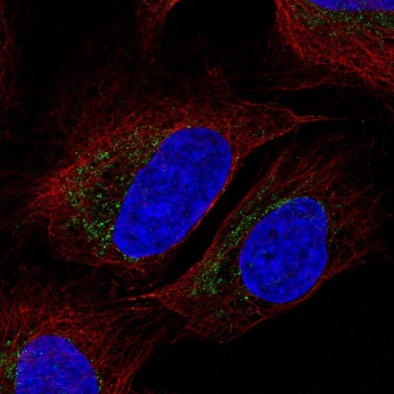
- Main image

- Experimental details
- Immunofluorescent staining of human cell line U-2 OS shows positivity in mitochondria.
- Sample type
- Human