Explore
 Validate
 Learn
Learn Western blot
Antibody data
- Antibody Data
- Antigen structure
- References [0]
- Comments [0]
- Validations
- Western blot [2]
- Immunocytochemistry [2]
- Immunohistochemistry [9]
Submit
Validation data
Reference
Comment
Report error
- Product number
- R31036 - Provider product page

- Provider
- NSJ Bioreagents
- Product name
- TFAM Antibody / mtTFA
- Antibody type
- Polyclonal
- Description
- This highly specific TFAM antibody is suitable for use in Western blot/Immunohistochemistry/Immunofluorescence applications with human samples.
- Reactivity
- Human
- Host
- Rabbit
- Conjugate
- Unconjugated
- Vial size
- 100 ug
- Concentration
- 0.5mg/ml if reconstituted with 0.2ml sterile DI water
- Storage
- After reconstitution, the TFAM antibody can be stored for up to one month at 4oC. For long-term, aliquot and store at -20oC. Avoid repeated freezing and thawing.
No comments: Submit comment
Supportive validation
- Submitted by
- NSJ Bioreagents (provider)
- Main image
- Experimental details
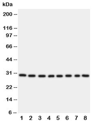
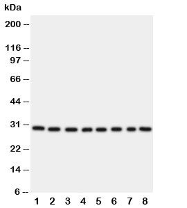
- Western blot testing of TFAM; Lane 1: HeLa; 2: Jurkat; 3: 293T; 4: A431; 5: Raji; 6: CEM; 7: HL-60; 8: HUT cell lysate. Expected molecular weight: 24~29 kDa.
- Submitted by
- NSJ Bioreagents (provider)
- Main image
- Experimental details
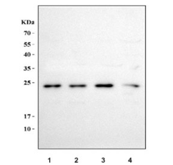
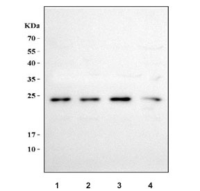
- Western blot testing of human 1) 293T, 2) K562, 3) Caco-2 and 4) Raji cell lysate with TFAM antibody. Expected molecular weight: 24-29 kDa.
Supportive validation
- Submitted by
- NSJ Bioreagents (provider)
- Main image
- Experimental details
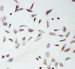
- ICC testing of TFAM antibody and HeLa cells

- Submitted by
- NSJ Bioreagents (provider)
- Main image

- Experimental details
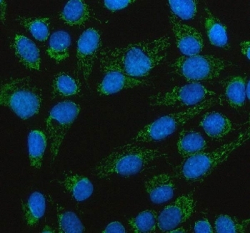
- Immunofluorescent staining of FFPE human U-2 OS cells with TFAM antibody (green) and DAPI nuclear stain (blue). HIER: steam section in pH6 citrate buffer for 20 min.
Supportive validation
- Submitted by
- NSJ Bioreagents (provider)
- Main image

- Experimental details
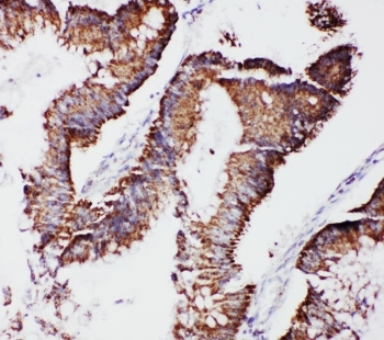
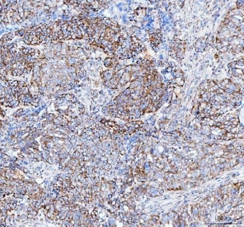
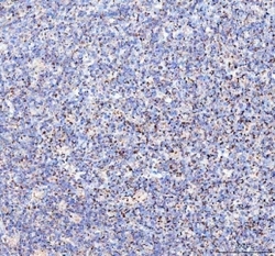
- IHC-P: TFAM antibody testing of human intestinal cancer tissue
- Submitted by
- NSJ Bioreagents (provider)
- Main image
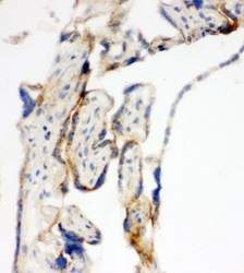
- Experimental details
- IHC-F testing of TFAM antibody and human placenta tissue

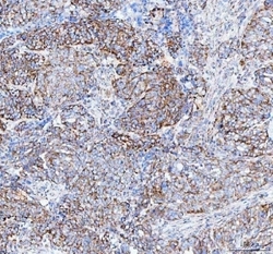
- Submitted by
- NSJ Bioreagents (provider)
- Main image
- Experimental details
- IHC staining of FFPE human liver cancer tissue with TFAM antibody. HIER: boil tissue sections in pH8 EDTA for 20 min and allow to cool before testing.
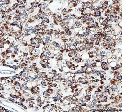
- Submitted by
- NSJ Bioreagents (provider)
- Main image
- Experimental details
- IHC staining of FFPE human ovarian serous adenocarcinoma tissue with TFAM antibody. HIER: boil tissue sections in pH8 EDTA for 20 min and allow to cool before testing.
- Submitted by
- NSJ Bioreagents (provider)
- Main image
- Experimental details
- IHC staining of FFPE human breast cancer tissue with TFAM antibody. HIER: boil tissue sections in pH8 EDTA for 20 min and allow to cool before testing.
- Submitted by
- NSJ Bioreagents (provider)
- Main image
- Experimental details
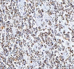
- IHC staining of FFPE human intestinal diffuse large B-cell lymphoma tissue with TFAM antibody. HIER: boil tissue sections in pH8 EDTA for 20 min and allow to cool before testing.
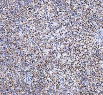
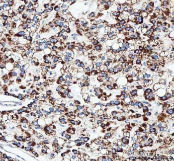
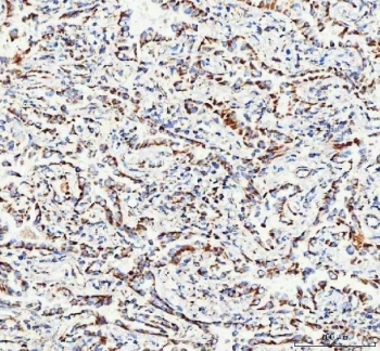
- Submitted by
- NSJ Bioreagents (provider)
- Main image
- Experimental details
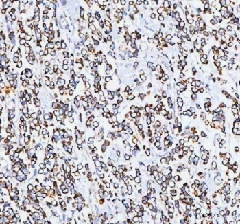
- IHC staining of FFPE human lung adenocarcinoma tissue with TFAM antibody. HIER: boil tissue sections in pH8 EDTA for 20 min and allow to cool before testing.
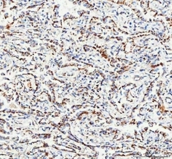
- Submitted by
- NSJ Bioreagents (provider)
- Main image

- Experimental details
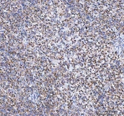
- IHC staining of FFPE human colorectal adenocarcinoma tissue with TFAM antibody. HIER: boil tissue sections in pH8 EDTA for 20 min and allow to cool before testing.
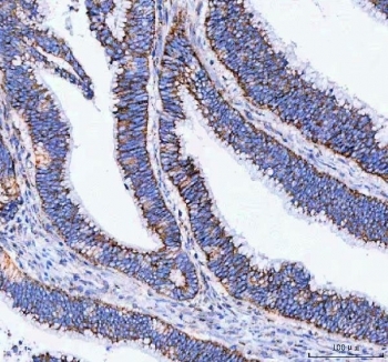
- Submitted by
- NSJ Bioreagents (provider)
- Main image
- Experimental details
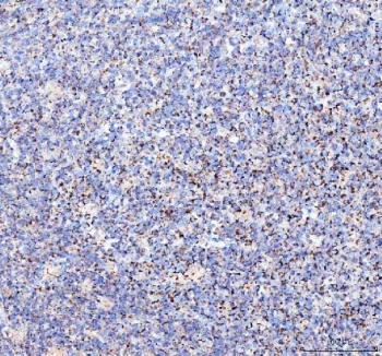
- IHC staining of FFPE human tonsil tissue with TFAM antibody. HIER: boil tissue sections in pH8 EDTA for 20 min and allow to cool before testing.