Explore
 Validate
 Learn
Learn Western blot
 ELISA
ELISAAntibody data
- Antibody Data
- Antigen structure
- References [0]
- Comments [0]
- Validations
- Western blot [3]
- Immunocytochemistry [1]
- Immunohistochemistry [1]
- Flow cytometry [1]
Submit
Validation data
Reference
Comment
Report error
- Product number
- F40574 - Provider product page

- Provider
- NSJ Bioreagents
- Product name
- TFAM Antibody
- Antibody type
- Polyclonal
- Description
- This highly specific TFAM antibody is suitable for use in Immunohistochemistry/Flow cytometry/Immunofluorescence/Western blot/ELISA applications with human samples.
- Reactivity
- Human
- Host
- Rabbit
- Conjugate
- Unconjugated
- Vial size
- 0.08 ml, 0.4 ml
- Concentration
- In 1X PBS, pH 7.4, with 0.09% sodium azide
- Storage
- Aliquot the TFAM antibody and store frozen at -20oC or colder. Avoid repeated freeze-thaw cycles.
No comments: Submit comment
Supportive validation
- Submitted by
- NSJ Bioreagents (provider)
- Main image
- Experimental details
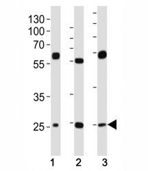
- Western blot analysis of lysate from (1) HeLa, (2) HepG2 and (3) U-2OS cell line using TFAM antibody at 1:1000. Expected molecular weight: 24~29 kDa.

- Submitted by
- NSJ Bioreagents (provider)
- Main image
- Experimental details
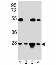
- TFAM antibody western blot analysis in (1) HeLa, (2) Jurkat, (3) K562, and (4) MCF-7 lysate; Expected molecular weight: 24~29 kDa.

- Submitted by
- NSJ Bioreagents (provider)
- Main image

- Experimental details
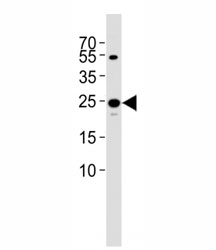
- Western blot analysis of lysate from 293T cell line using TFAM antibody diluted at 1:1000. Expected molecular weight: 24~29 kDa.
Supportive validation
- Submitted by
- NSJ Bioreagents (provider)
- Main image
- Experimental details
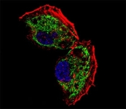
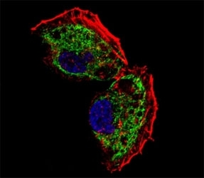
- Fluorescent confocal image of NCI-H460 cell stained with TFAM antibody at 1:25. TFAM immunoreactivity is localized to mitochondrion.
Supportive validation
- Submitted by
- NSJ Bioreagents (provider)
- Main image
- Experimental details
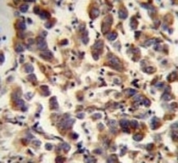
- TFAM antibody immunohistochemistry analysis in formalin fixed and paraffin embedded human testis carcinoma.

Supportive validation
- Submitted by
- NSJ Bioreagents (provider)
- Main image
- Experimental details
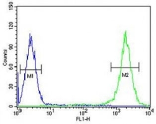
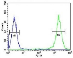
- TFAM antibody flow cytometric analysis of K562 cells (right histogram) compared to a negative control (left histogram). FITC-conjugated goat-anti-rabbit secondary Ab was used for the analysis.