 Explore
 Validate
 Learn
Learn Western blot
Antibody data
- Antibody Data
- Antigen structure
- References [4]
- Comments [0]
- Validations
- Western blot [3]
- Immunocytochemistry [1]
- Immunohistochemistry [2]
- Flow cytometry [2]
- Other assay [2]
Submit
Validation data
Reference
Comment
Report error
- Product number
- PA5-23776 - Provider product page

- Provider
- Invitrogen Antibodies
- Product name
- TFAM Polyclonal Antibody
- Antibody type
- Polyclonal
- Antigen
- Synthetic peptide
- Reactivity
- Human
- Host
- Rabbit
- Isotype
- IgG
- Vial size
- 400 μL
- Concentration
- 0.38 mg/mL
- Storage
- Store at 4°C short term. For long term storage, store at -20°C, avoiding freeze/thaw cycles.
Submitted references Prevention of HIV-1 TAT Protein-Induced Peripheral Neuropathy and Mitochondrial Disruption by the Antimuscarinic Pirenzepine.
Mitochondrial biogenesis is altered in HIV+ brains exposed to ART: Implications for therapeutic targeting of astroglia.
Tenofovir disoproxil fumarate induces peripheral neuropathy and alters inflammation and mitochondrial biogenesis in the brains of mice.
SYBR Gold dye enables preferential labelling of mitochondrial nucleoids and their time-lapse imaging by structured illumination microscopy.
Han MM, Frizzi KE, Ellis RJ, Calcutt NA, Fields JA
Frontiers in neurology 2021;12:663373
Frontiers in neurology 2021;12:663373
Mitochondrial biogenesis is altered in HIV+ brains exposed to ART: Implications for therapeutic targeting of astroglia.
Swinton MK, Carson A, Telese F, Sanchez AB, Soontornniyomkij B, Rad L, Batki I, Quintanilla B, Pérez-Santiago J, Achim CL, Letendre S, Ellis RJ, Grant I, Murphy AN, Fields JA
Neurobiology of disease 2019 Oct;130:104502
Neurobiology of disease 2019 Oct;130:104502
Tenofovir disoproxil fumarate induces peripheral neuropathy and alters inflammation and mitochondrial biogenesis in the brains of mice.
Fields JA, Swinton MK, Carson A, Soontornniyomkij B, Lindsay C, Han MM, Frizzi K, Sambhwani S, Murphy A, Achim CL, Ellis RJ, Calcutt NA
Scientific reports 2019 Nov 20;9(1):17158
Scientific reports 2019 Nov 20;9(1):17158
SYBR Gold dye enables preferential labelling of mitochondrial nucleoids and their time-lapse imaging by structured illumination microscopy.
Jevtic V, Kindle P, Avilov SV
PloS one 2018;13(9):e0203956
PloS one 2018;13(9):e0203956
No comments: Submit comment
Supportive validation
- Submitted by
- Invitrogen Antibodies (provider)
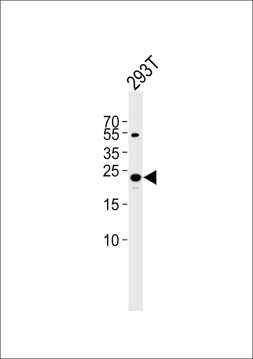
- Main image

- Experimental details
- Western blot analysis of TFAM in 293T cell line. Samples were incubated with TFAM polyclonal antibody (Product # PA5-23776) using a dilution of 1:1,000 followed by goat anti-rabbit IgG H&L (HRP) at a dilution of 1:5,000. Lysate at 35 µg.
- Submitted by
- Invitrogen Antibodies (provider)
- Main image
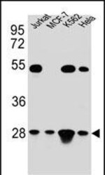
- Experimental details
- Western blot analysis of TFAM in Hela, Jurkat, K562, MCF-7 cell line lysates. Samples were incubated with TFAM polyclonal antibody (Product # PA5-23776). Lysates: 35 µg/lane. TFAM protein (arrow).

- Submitted by
- Invitrogen Antibodies (provider)
- Main image
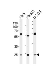
- Experimental details
- Western blot analysis of TFAM in lysates from Hela, HepG2, U-2OS cell line (from left to right). Samples were incubated with TFAM polyclonal antibody (Product # PA5-23776) using a dilution of 1:1,000 followed by goat anti-rabbit IgG H&L (HRP) at a dilution of 1:10,000. Lysates at 20 µg per lane.

Supportive validation
- Submitted by
- Invitrogen Antibodies (provider)
- Main image
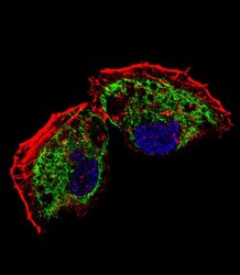
- Experimental details
- Immunocytochemistry analysis of TFAM in NCI-H460 cells. Samples were incubated with TFAM polyclonal antibody (Product # PA5-23776) using a dilution of 1:25 for 1 h at 37°C followed by Alexa Fluor® 488 conjugated donkey anti-rabbit at a dilution of 1:400 for 50 min at 37°C. Cells were fixed with 4% PFA (20 min) and permeabilized with Triton X-100 (0.1%, 10 min). Cytoplasmic actin was counterstained with Alexa Fluor® 555 (red) conjugated Phalloidin (7 units/mL, 1 h at 37°C). Nuclei were counterstained with DAPI (blue) (10 µg/mL, 10 min). TFAM immunoreactivity is localized to mitochondrion significantly.

Supportive validation
- Submitted by
- Invitrogen Antibodies (provider)
- Main image

- Experimental details
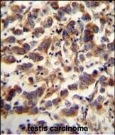
- Immunohistochemistry analysis of TFAM in formalin fixed and paraffin embedded human testis carcinoma. Samples were incubated with TFAM polyclonal antibody (Product # PA5-23776) followed by peroxidase conjugation of the secondary antibody and DAB staining. This data demonstrates the use of this antibody for immunohistochemistry. Clinical relevance has not been evaluated.
- Submitted by
- Invitrogen Antibodies (provider)
- Main image
- Experimental details
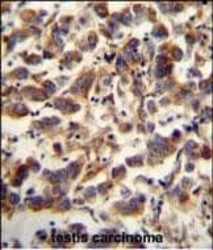
- Immunohistochemistry analysis of TFAM in formalin fixed and paraffin embedded human testis carcinoma. Samples were incubated with TFAM polyclonal antibody (Product # PA5-23776) followed by peroxidase conjugation of the secondary antibody and DAB staining. This data demonstrates the use of this antibody for immunohistochemistry. Clinical relevance has not been evaluated.

Supportive validation
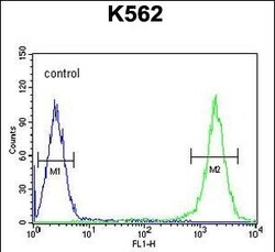
- Submitted by
- Invitrogen Antibodies (provider)
- Main image
- Experimental details
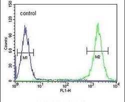
- Flow cytometry analysis of K562 cells using a TFAM polyclonal antibody (Product # PA5-23776) (right) compared to a negative control cell (left) at a dilution of 1:10-50, followed by a FITC-conjugated goat anti-rabbit antibody
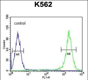
- Submitted by
- Invitrogen Antibodies (provider)
- Main image
- Experimental details
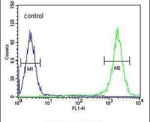
- Flow cytometry of TFAM in K562 cells (right histogram). Samples were incubated with TFAM polyclonal antibody (Product # PA5-23776) followed by FITC-conjugated goat-anti-rabbit secondary antibody. Negative control cell (left histogram).
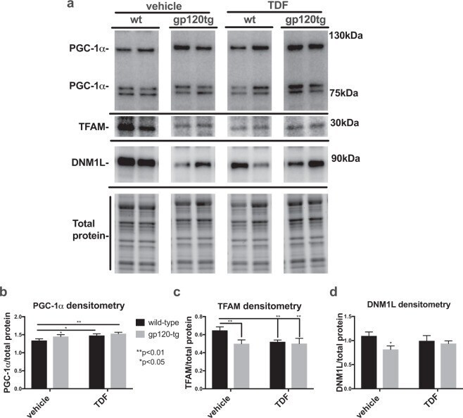
Supportive validation
- Submitted by
- Invitrogen Antibodies (provider)
- Main image
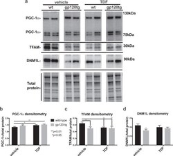
- Experimental details
- Figure 1 PGC-1alpha protein levels are increased and TFAM and DNM1L protein levels are decreased in brain lysates from wt and gp120-tg mice administered Tenofovir disproxil fumarate. ( a ) Immunoblot for PGC-1alpha, TFAM, and DNM1L using whole lysates from hippocampal region. ( b--d ) Densitometry for quantification of PGC-1alpha, and TFAM, and DNM1L normalized to total protein levels. Significance (p < 0.05) was determined by two-way ANOVA (wt, n = 8; wt + TDF, n = 5; gp120-tg, n = 7; gp120-tg, n = 7).
- Submitted by
- Invitrogen Antibodies (provider)
- Main image
- Experimental details
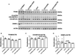
- Figure 4 Mitochondrial proteins in mouse sciatic nerve. (A) Representative immunoblot for TFAM, proteins of oxidative phosphorylation complexes III, IV, and V, DNM1L and ACTB. (B) Densitometric analysis of TFAM normalized to ACTB. (C) Densitometric analysis of complex IV(MTC01 sub-unit) protein normalized to ACTB. (D) Densitometric analyses of DNM1L protein normalized to ACTB. Data are mean + SEM of N = 3 for C57Bl/6J group and N = 4/group for remaining groups. Statistical analyses by unpaired test or one-way ANOVA with Tukey's post-hoc test.
