Explore
 Validate
 Learn
Learn Western blot
Antibody data
- Antibody Data
- Antigen structure
- References [0]
- Comments [0]
- Validations
- Western blot [3]
- Immunocytochemistry [3]
- Immunohistochemistry [2]
- Other assay [1]
Submit
Validation data
Reference
Comment
Report error
- Product number
- PA5-27865 - Provider product page

- Provider
- Invitrogen Antibodies
- Product name
- TFAM Polyclonal Antibody
- Antibody type
- Polyclonal
- Antigen
- Recombinant protein fragment
- Description
- Recommended positive controls: 293T, A431, HeLa, HepG2.
- Concentration
- 1.23 mg/mL
No comments: Submit comment
Supportive validation
- Submitted by
- Invitrogen Antibodies (provider)
- Main image
- Experimental details
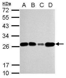
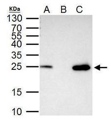
- Western Blot using TFAM Polyclonal Antibody (Product # PA5-27865). Sample (30 µg of whole cell lysate). Lane A: 293T. Lane B: A431. Lane C: HeLa. Lane D: HepG2. 12% SDS PAGE. TFAM Polyclonal Antibody (Product # PA5-27865) diluted at 1:5,000. The HRP-conjugated anti-rabbit IgG antibody was used to detect the primary antibody.
- Submitted by
- Invitrogen Antibodies (provider)
- Main image
- Experimental details
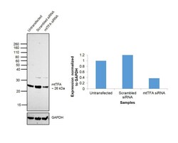
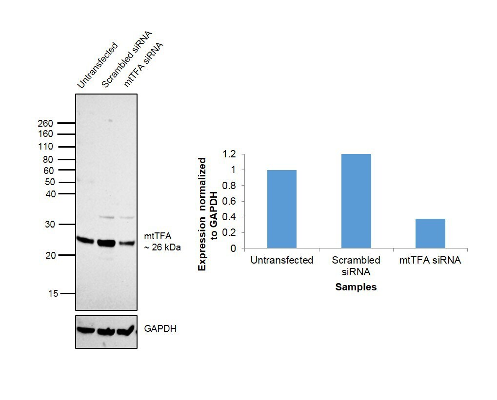
- Knockdown of mtTFA was achieved by transfecting HeLa cells with mtTFA specific siRNAs (Silencer® select Product # s14000, s14001). Western blot analysis (Fig. a) was performed membrane enriched extracts from the HeLa knockdown cells (lane 3), non-specific scrambled siRNA transfected cells (lane 2) and untransfected cells (lane 1). The blot was probed with mtTFA Polyclonal Antibody (Product # PA5-27865, 1:5000 dilution) and Goat anti-Rabbit IgG (H+L) Superclonal™ Recombinant Secondary Antibody, HRP (Product # A27036, 1:4000 dilution). Densitometric analysis of this western blot is shown in histogram (Fig. b). Decrease in signal upon siRNA mediated knock down confirms that antibody is specific to mtTFA.
- Submitted by
- Invitrogen Antibodies (provider)
- Main image
- Experimental details
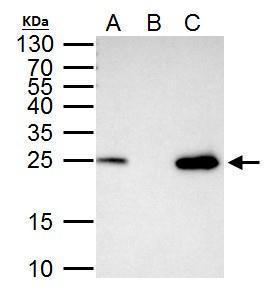
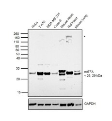
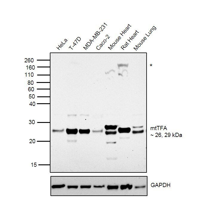
- Western blot was performed using Anti-mtTFA Rabbit Polyclonal Antibody (Product # PA5-27865) and 26 kDa, 29 kDa bands corresponding to mtTFA were observed in cell lines and tissues tested along with an uncharacterized band (*) in Rat Heart. Membrane enriched extracts (30 µg lysate) of HeLa (Lane 1), T-47D (Lane 2), MDA-MB-231 Lung (Lane 3), Caco-2 (Lane 4), Mouse Heart (Lane 5), Rat Heart (Lane 6) and Mouse Lung (Lane 7) were electrophoresed using NuPAGE™ 12% Bis-Tris Protein Gel (Product # NP0342BOX). Resolved proteins were then transferred onto a nitrocellulose membrane (Product # IB23001) by iBlot® 2 Dry Blotting System (Product # IB21001). The blot was probed with the primary antibody (1:5000 dilution) and detected by chemiluminescence with Goat anti-Rabbit IgG (H+L) Superclonal™ Recombinant Secondary Antibody, HRP (Product # A27036, 1:4000 dilution) using the iBright FL 1000 (Product # A32752). Chemiluminescent detection was performed using Novex® ECL Chemiluminescent Substrate Reagent Kit (Product # WP20005).
Supportive validation
- Submitted by
- Invitrogen Antibodies (provider)
- Main image
- Experimental details
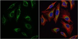
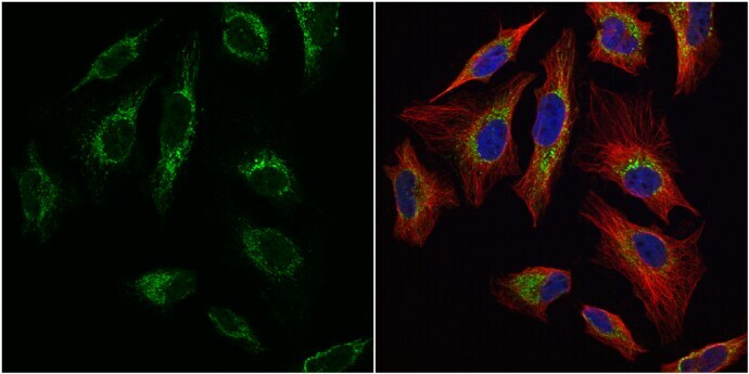
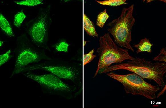
- Immunocytochemistry-Immunofluorescence analysis of TFAM was performed in HeLa cells fixed in 4% paraformaldehyde at RT for 15 min. Green: TFAM Polyclonal Antibody (Product # PA5-27865) diluted at 1:1000. Red: alpha Tubulin, a cytoskeleton marker. Blue: Hoechst 33342 staining.
- Submitted by
- Invitrogen Antibodies (provider)
- Main image
- Experimental details
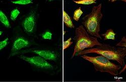
- TFAM Polyclonal Antibody detects mtTFA protein at mito by immunofluorescent analysis. Sample: HeLa cells were fixed in 4% paraformaldehyde at RT for 15 min. Green: mtTFA stained by TFAM Polyclonal Antibody (Product # PA5-27865) diluted at 1:500. Red: alpha Tubulin, a cytoskeleton marker, stained by alpha Tubulin Polyclonal Antibody [GT114] (Product # MA5-31466) diluted at 1:1,000. Blue: Fluoroshield with DAPI .
- Submitted by
- Invitrogen Antibodies (provider)
- Main image
- Experimental details
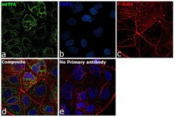
- Immunofluorescence analysis of mtTFA was performed using Caco-2 cells. The cells were fixed with 4% paraformaldehyde for 10 minutes, permeabilized with 0.1% Triton™ X-100 for 15 minutes, and blocked with 2% BSA for 1 hour at room temperature. The cells were labeled with mtTFA Rabbit Polyclonal Antibody (Product # PA5-27865) at 1:500 dilution in 0.1% BSA and incubated overnight at 4 degree and then labeled with Goat anti-Rabbit IgG (H+L) Highly Cross-Adsorbed Secondary Antibody, Alexa Fluor Plus 488 (Product # A32731) at a dilution of 1:2000 for 45 minutes at room temperature (Panel a: green). Nuclei (Panel b: blue) were stained with ProLong™ Diamond Antifade Mountant with DAPI (Product # P36962). F-actin (Panel c: red) was stained with Rhodamine Phalloidin (Product # R415, 1:300). Panel d represents the composite image showing mitochondrial pattern of mtTFA. Panel e represents control cells with no primary antibody to assess background. The images were captured at 60X magnification.

Supportive validation
- Submitted by
- Invitrogen Antibodies (provider)
- Main image
- Experimental details
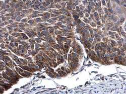
- Immunohistochemistry (Paraffin) analysis of TFAM was performed in paraffin-embedded human esophagus cancer tissue using TFAM Polyclonal Antibody (Product # PA5-27865) at a dilution of 1:500.

- Submitted by
- Invitrogen Antibodies (provider)
- Main image

- Experimental details
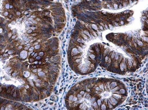
- Immunohistochemistry (Paraffin) analysis of TFAM was performed in paraffin-embedded human colon cancer tissue using TFAM Polyclonal Antibody (Product # PA5-27865) at a dilution of 1:500.
Supportive validation
- Submitted by
- Invitrogen Antibodies (provider)
- Main image
- Experimental details
- TFAM Polyclonal Antibody immunoprecipitates mtTFA protein in IP experiments. IP Sample: 293T whole cell lysate/extract A. 40 µg 293T whole cell lysate/extract B. Control with 2 µg of preimmune rabbit IgG C. Immunoprecipitation of mtTFA protein by 2 µg of TFAM Polyclonal Antibody (Product # PA5-27865) 15% SDS-PAGE The immunoprecipitated mtTFA protein was detected by TFAM Polyclonal Antibody (Product # PA5-27865) diluted at 1:1,000.