Explore
 Validate
 Learn
Learn Western blot
Antibody data
- Antibody Data
- Antigen structure
- References [3]
- Comments [0]
- Validations
- Western blot [1]
- Immunocytochemistry [1]
- Immunoprecipitation [1]
- Immunohistochemistry [2]
Submit
Validation data
Reference
Comment
Report error
- Product number
- GTX103231 - Provider product page

- Provider
- GeneTex
- Proper citation
- GeneTex Cat#GTX103231, RRID:AB_11176720
- Product name
- mtTFA antibody
- Antibody type
- Polyclonal
- Reactivity
- Human, Mouse
- Host
- Rabbit
Submitted references Neurotoxicity of cytarabine (Ara-C) in dorsal root ganglion neurons originates from impediment of mtDNA synthesis and compromise of mitochondrial function.
Cisplatin Toxicity in Dorsal Root Ganglion Neurons Is Relieved by Meclizine via Diminution of Mitochondrial Compromise and Improved Clearance of DNA Damage.
Inhibition of cyclophilin D by cyclosporin A promotes retinal ganglion cell survival by preventing mitochondrial alteration in ischemic injury.
Zhuo M, Gorgun MF, Englander EW
Free radical biology & medicine 2018 Jun;121:9-19
Free radical biology & medicine 2018 Jun;121:9-19
Cisplatin Toxicity in Dorsal Root Ganglion Neurons Is Relieved by Meclizine via Diminution of Mitochondrial Compromise and Improved Clearance of DNA Damage.
Gorgun MF, Zhuo M, Englander EW
Molecular neurobiology 2017 Dec;54(10):7883-7895
Molecular neurobiology 2017 Dec;54(10):7883-7895
Inhibition of cyclophilin D by cyclosporin A promotes retinal ganglion cell survival by preventing mitochondrial alteration in ischemic injury.
Kim SY, Shim MS, Kim KY, Weinreb RN, Wheeler LA, Ju WK
Cell death & disease 2014 Mar 6;5:e1105
Cell death & disease 2014 Mar 6;5:e1105
No comments: Submit comment
Supportive validation
- Submitted by
- GeneTex (provider)
- Main image
- Experimental details
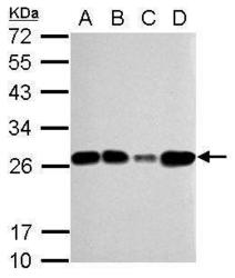
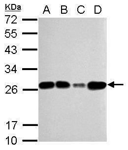
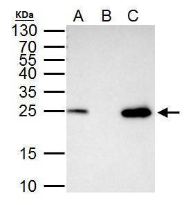
- Sample (30 ?g of whole cell lysate) A: 293T B: A431 C: HeLa D: HepG2 12% SDS PAGE GTX103231 diluted at 1:5000 The HRP-conjugated anti-rabbit IgG antibody (GTX213110-01) was used to detect the primary antibody.
Supportive validation
- Submitted by
- GeneTex (provider)
- Main image

- Experimental details
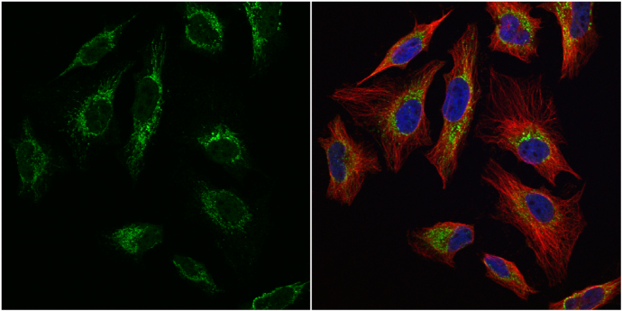
- mtTFA antibody detects mtTFA protein at mitochondria by immunofluorescent analysis.Sample: HeLa cells were fixed in 4% paraformaldehyde at RT for 15 min.Green: mtTFA protein stained by mtTFA antibody (GTX103231) diluted at 1:1000.Red: alpha Tubulin, a cytoskeleton marker, stained by alpha Tubulin antibody [B-5-1-2] (GTX11304) diluted at 1:10000.Blue: Hoechst 33342 staining.
Supportive validation
- Submitted by
- GeneTex (provider)
- Main image
- Experimental details
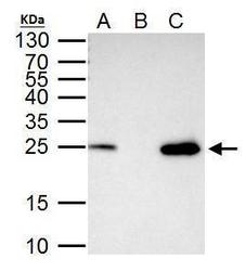
- mtTFA antibody immunoprecipitates mtTFA protein in IP experiments. IP Sample: 293T whole cell lysate/extract A. 40 £gg 293T whole cell lysate/extract B. Control with 2 £gg of preimmune rabbit IgG C. Immunoprecipitation of mtTFA protein by 2 £gg of mtTFA antibody (GTX103231) 15% SDS-PAGE The immunoprecipitated mtTFA protein was detected by mtTFA antibody (GTX103231) diluted at 1:1000. EasyBlot anti-rabbit IgG (GTX221666-01) was used as a secondary reagent.
Supportive validation
- Submitted by
- GeneTex (provider)
- Main image
- Experimental details
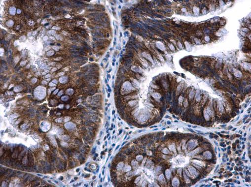
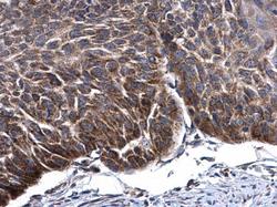
- mtTFA antibody detects mtTFA protein at cytoplasm in human esophagus cancer by immunohistochemical analysis. Sample: Paraffin-embedded human esophagus cancer. mtTFA antibody (GTX103231) diluted at 1:500.

- Submitted by
- GeneTex (provider)
- Main image

- Experimental details
- mtTFA antibody detects mtTFA protein at cytoplasm in human colon cancer by immunohistochemical analysis. Sample: Paraffin-embedded human colon cancer. mtTFA antibody (GTX103231) diluted at 1:500.