 Explore
 Validate
 Learn
Learn Western blot
Antibody data
- Antibody Data
- Antigen structure
- References [1]
- Comments [0]
- Validations
- Western blot [1]
- Immunocytochemistry [1]
- Immunohistochemistry [1]
Submit
Validation data
Reference
Comment
Report error
- Product number
- AF5919 - Provider product page

- Provider
- R&D Systems
- Product name
- Mouse/Rat ADNP Antibody
- Antibody type
- Polyclonal
- Description
- Immunogen affinity purified. Detects mouse and rat ADNP in direct ELISAs and Western blots. In direct ELISAs, approximately 30% cross-reactivity with recombinant human ADNP is observed.
- Reactivity
- Mouse, Rat
- Host
- Goat
- Conjugate
- Unconjugated
- Antigen sequence
Q9H2P0- Isotype
- IgG
- Vial size
- 100 ug
- Concentration
- LYOPH
- Storage
- Use a manual defrost freezer and avoid repeated freeze-thaw cycles. 12 months from date of receipt, -20 to -70 °C as supplied. 1 month, 2 to 8 °C under sterile conditions after reconstitution. 6 months, -20 to -70 °C under sterile conditions after reconstitution.
Submitted references Novel evolutionary-conserved role for the activity-dependent neuroprotective protein (ADNP) family that is important for erythropoiesis.
Dresner E, Malishkevich A, Arviv C, Leibman Barak S, Alon S, Ofir R, Gothilf Y, Gozes I
The Journal of biological chemistry 2012 Nov 23;287(48):40173-85
The Journal of biological chemistry 2012 Nov 23;287(48):40173-85
No comments: Submit comment
Supportive validation
- Submitted by
- R&D Systems (provider)
- Main image

- Experimental details
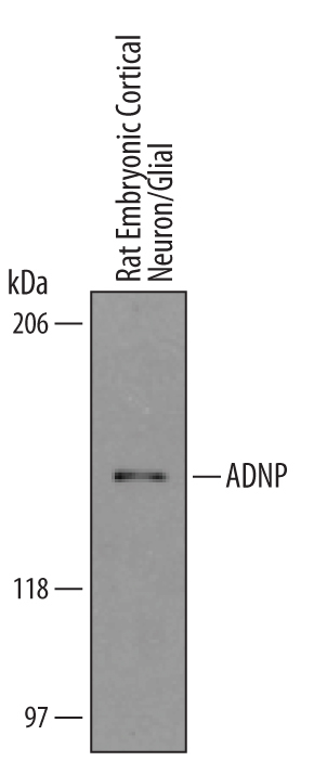
- Detection of Mouse/Rat ADNP by Western Blot. Western blot shows lysates of embryonic rat cortical neuron/ glial cells. PVDF membrane was probed with 1 µg/mL of Goat Anti-Mouse/Rat ADNP Antigen Affinity-purified Polyclonal Antibody (Catalog # AF5919) followed by HRP-conjugated Anti-Goat IgG Secondary Antibody (Catalog # HAF019). A specific band was detected for ADNP at approximately 150 kDa (as indicated). This experiment was conducted under reducing conditions and using Immunoblot Buffer Group 1.
Supportive validation
- Submitted by
- R&D Systems (provider)
- Main image

- Experimental details
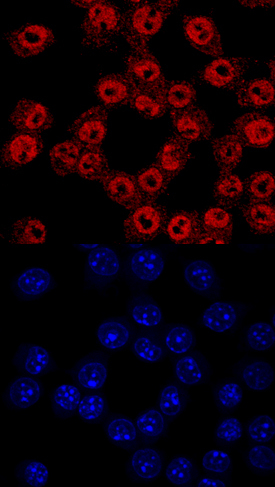
- ADNP in RAW 264.7 Mouse Cell Line. ADNP was detected in immersion fixed RAW 264.7 mouse monocyte/macrophage cell line using Goat Anti-Mouse/Rat ADNP Antigen Affinity-purified Polyclonal Antibody (Catalog # AF5919) at 10 µg/mL for 3 hours at room temperature. Cells were stained using the NorthernLights™ 557-conjugated Anti-Goat IgG Secondary Antibody (red, upper panel; Catalog # NL001) and counterstained with DAPI (blue, lower panel). Specific staining was localized to nuclei. View our protocol for Fluorescent ICC Staining of Cells on Coverslips.
Supportive validation
- Submitted by
- R&D Systems (provider)
- Main image
- Experimental details
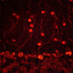
- ADNP in Mouse Brain. ADNP was detected in perfusion fixed frozen sections of mouse brain (cerebellum) using Goat Anti-Mouse/Rat ADNP Antigen Affinity-purified Polyclonal Antibody (Catalog # AF5919) at 1.7 µg/mL overnight at 4 °C. Tissue was stained using the NorthernLights™ 557-conjugated Anti-Goat IgG Secondary Antibody (red; Catalog # NL001). Specific staining was localized to Purkinje cells and neurons in molecular cell layer. View our protocol for Fluorescent IHC Staining of Frozen Tissue Sections.
