Explore
 Validate
 Learn
Learn Western blot
 Immunocytochemistry
Antibody data
- Antibody Data
- Antigen structure
- References [1]
- Comments [0]
- Validations
- Immunocytochemistry [1]
- Other assay [1]
Submit
Validation data
Reference
Comment
Report error
- Product number
- 703166 - Provider product page

- Provider
- Invitrogen Antibodies
- Product name
- ZBP1 Recombinant Rabbit Monoclonal Antibody (5H15L56)
- Antibody type
- Monoclonal
- Antigen
- Other
- Description
- This antibody is predicted to react with Mouse, Rat, Bovine.
- Reactivity
- Human
- Host
- Rabbit
- Isotype
- IgG
- Antibody clone number
- 5H15L56
- Vial size
- 100 μg
- Concentration
- 0.5 mg/mL
- Storage
- Store at 4°C short term. For long term storage, store at -20°C, avoiding freeze/thaw cycles.
Submitted references SARS-CoV-2 Infects Primary Neurons from Human ACE2 Expressing Mice and Upregulates Genes Involved in the Inflammatory and Necroptotic Pathways.
Rothan HA, Kumari P, Stone S, Natekar JP, Arora K, Auroni TT, Kumar M
Pathogens (Basel, Switzerland) 2022 Feb 17;11(2)
Pathogens (Basel, Switzerland) 2022 Feb 17;11(2)
No comments: Submit comment
Supportive validation
- Submitted by
- Invitrogen Antibodies (provider)
- Main image
- Experimental details
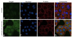
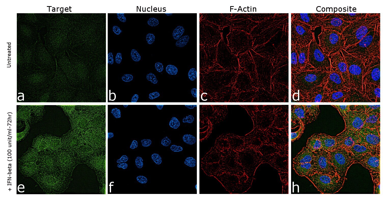
- For immunofluorescence analysis, A549 cells were fixed and permeabilized for detection of endogenous ZBP1 using Anti-ZBP1 Recombinant Rabbit Monoclonal Antibody (Product # 703166, 1:100 dilution) and labeled with Goat anti-Rabbit IgG (H+L) Highly Cross-Adsorbed Secondary Antibody, Alexa Fluor Plus 488 conjugate (Product # A32731, 1:2000). Nuclei (blue) were stained using ProLong™ Diamond Antifade Mountant with DAPI (Product # P36962) and cytoskeletal F-actin (red) staining using Rhodamine Phalloidin (Product # R415, 1:300) Panel a-d) shows representative untreated A549 cells that were stained for detection and localization of ZBP1 protein (green) while panel e-h) demonstrates upregulation of cytoplasmic localization of ZBP1 upon treatment with IFN-beta 100 unit/mL for 72hrs. The images were captured at 60X magnification.
Supportive validation
- Submitted by
- Invitrogen Antibodies (provider)
- Main image
- Experimental details
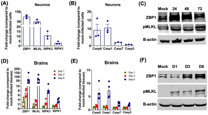
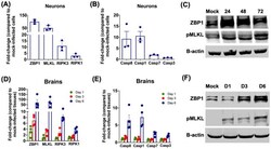
- Figure 4 mRNA and protein levels of genes involved in cell-death pathways in primary mouse neurons and mouse brains. ( A , B ) The hACE2 neurons were infected with SARS-CoV-2 or mock-infected at a MOI of 0.1. At 48 h after infection, cell pellets were collected, and total RNA was extracted. qRT-PCR was conducted to determine the fold-change in ( A ) ZBP1, MLKL, RIPK3 and RIPK1, and ( B ) Caspase 8, Caspase 1, Caspase 7 and Caspase 8 mRNA levels. Data for each sample was normalized to the value for GAPDH and expressed as the relative fold increase compared to mock-infected controls. Data represent the mean +- SEM of three independent infection experiments conducted in duplicate. Each data point represents an independent experiment. ( C ) The hACE2 neurons were infected with SARS-CoV-2 or mock-infected at a MOI of 0.1. At 24, 48 and 72 h, cell pellets were collected, and total protein was extracted. Protein was blotted with ZBP1, pMLKL or beta-actin antibodies. Data are representative of three independent experiments. ( D , E ) K18-hACE2 mice were inoculated with SARS-CoV-2 or PBS via the intranasal route. Brains were harvested after extensive perfusion at days 1, 3 and 6 after infection and RNA was extracted. qRT-PCR was used to determine the mRNA levels of ( D ) ZBP1, MLKL, RIPK3 and RIPK1, and ( E ) Caspase 8, Caspase 1, Caspase 7 and Caspase 8. After normalizing individual sample to GAPDH level, the fold change in infected tissues compared to mock-infected controls was dete