 Explore
 Validate
 Learn
LearnPA5-20014
antibody from Invitrogen Antibodies
Targeting: NLRC4
CARD12, CLAN, CLAN1, CLANA, CLANB, CLANC, CLAND, CLR2.1, ipaf
 Western blot
Antibody data
- Antibody Data
- Antigen structure
- References [0]
- Comments [0]
- Validations
- Western blot [3]
- Immunocytochemistry [3]
- Immunohistochemistry [1]
Submit
Validation data
Reference
Comment
Report error
- Product number
- PA5-20014 - Provider product page

- Provider
- Invitrogen Antibodies
- Product name
- NLRC4 Polyclonal Antibody
- Antibody type
- Polyclonal
- Antigen
- Synthetic peptide
- Description
- In Western blot applications, this antibody detects a band at ~110kDa. A suggested positive control is human peripheral blood leukocyte (pbl) lysate.
- Concentration
- 1 mg/mL
No comments: Submit comment
Supportive validation
- Submitted by
- Invitrogen Antibodies (provider)
- Main image

- Experimental details
- Western Blot analysis of Ipaf in human PBL lysate with NLRC4 Polyclonal Antibody (Product # PA5-20014) at 0.5 (lane A), 1 (lane B), and 2 (lane C) µg/mL, respectively.

- Submitted by
- Invitrogen Antibodies (provider)
- Main image
- Experimental details
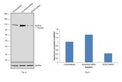
- Knockdown of NLR family CARD domain-containing protein 4 was achieved by transfecting THP-1 with NLRC4 specific siRNAs (Silencer® select Product # s33828, s33829). Western blot analysis (Fig. a) was performed using whole cell extracts from the NLRC4 knockdown cells (lane 3), non-targeting scrambled siRNA transfected cells (lane 2) and untransfected cells (lane 1). The blot was probed with NLRC4 Polyclonal Antibody (Product # PA5-20014, 1.5 µg/mL dilution) and Goat anti-Rabbit IgG (H+L) Superclonal™ Recombinant Secondary Antibody, HRP (Product # A27036, 1:10000 dilution). Densitometric analysis of this western blot is shown in histogram (Fig. b). Decrease in signal upon siRNA mediated knock down confirms that antibody is specific to NLR family CARD domain-containing protein 4.

- Submitted by
- Invitrogen Antibodies (provider)
- Main image
- Experimental details
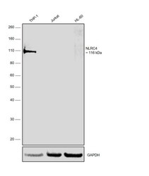
- Western blot was performed using Anti-NLRC4 Polyclonal Antibody (Product # PA5-20014) and a 116 kDa band corresponding to NLRC4 was observed in THP-1 and not in Jurkat and HL-60 which are reported low expressing for the same. Whole cell extracts (40 µg lysate) of THP-1 (Lane 1), Jurkat (Lane 2), HL-60 (Lane 3) were electrophoresed using NuPAGE™ 4-12% Bis-Tris Protein Gel (Product # NP0321BOX). Resolved proteins were then transferred onto a nitrocellulose membrane (Product # IB23001) by iBlot® 2 Dry Blotting System (Product # IB21001). The blot was probed with the primary antibody (1.5 µg/mL dilution) and detected by chemiluminescence with Goat anti-Rabbit IgG (H+L) Superclonal™ Recombinant Secondary Antibody, HRP (Product # A27036, 1:10000 dilution) using the iBright FL 1000 (Product # A32752). Chemiluminescent detection was performed using SuperSignal™ West Atto Ultimate Sensitivity Substrate (Product # A38556).

Supportive validation
- Submitted by
- Invitrogen Antibodies (provider)
- Main image

- Experimental details
- Immunocytochemistry of Ipaf in THP-1 cells with NLRC4 Polyclonal Antibody (Product # PA5-20014) at 10 µg/mL.

- Submitted by
- Invitrogen Antibodies (provider)
- Main image

- Experimental details
- Immunofluorescence of Ipaf in THP1 cells with NLRC4 Polyclonal Antibody (Product # PA5-20014) at 10 µg/mL.

- Submitted by
- Invitrogen Antibodies (provider)
- Main image

- Experimental details
- Immunofluorescence analysis of NLRC4 was performed using THP-1 and Jurkat cells. The cells were fixed with 4% paraformaldehyde for 10 minutes, permeabilized with 0.1% Triton™ X-100 for 10 minutes, and blocked with 2% BSA for 10 minutes at room temperature. The cells were labeled with NLRC4 Polyclonal Antibody (Product # PA5-20014) at 10 µg in 0.1% BSA, incubated at 4 degree celsius overnight and then labeled with Donkey anti-Rabbit IgG (H+L) Highly Cross-Adsorbed Secondary Antibody, Alexa Fluor Plus 488 (Product # A32790), (1:2500 dilution), for 45 minutes at room temperature (Panel a: Green). Nuclei (Panel b: Blue) were stained with ProLong™ Diamond Antifade Mountant with DAPI (Product # P36962). F-actin (Panel c: Red) was stained with Rhodamine Phalloidin (Product # R415, 1:300). Panel d represents the merged image showing cytoplasmic localization of NLRC4 in THP-1 and panel e represents Jurkat cells showing no signal for the same. Panel f represents control cells with no primary antibody to assess background. The images were captured at 60X magnification.

Supportive validation
- Submitted by
- Invitrogen Antibodies (provider)
- Main image

- Experimental details
- Immunocytochemistry staining of THP-1 cells using a Ipaf polyclonal antibody (Product # PA5-20014) at a 10 µg/mL dilution.
