Explore
 Validate
 Learn
Learn Western blot
 Immunocytochemistry
Antibody data
- Antibody Data
- Antigen structure
- References [1]
- Comments [0]
- Validations
- Immunocytochemistry [1]
- Immunohistochemistry [1]
Submit
Validation data
Reference
Comment
Report error
- Product number
- HPA019051 - Provider product page

- Provider
- Atlas Antibodies
- Proper citation
- Atlas Antibodies Cat#HPA019051, RRID:AB_1846164
- Product name
- Anti-CCDC6
- Antibody type
- Polyclonal
- Description
- Polyclonal Antibody against Human CCDC6, Gene description: coiled-coil domain containing 6, Alternative Gene Names: D10S170, H4, PTC, TPC, TST1, Validated applications: ICC, IHC, WB, Uniprot ID: Q16204, Storage: Store at +4°C for short term storage. Long time storage is recommended at -20°C.
- Reactivity
- Human, Mouse, Rat
- Host
- Rabbit
- Conjugate
- Unconjugated
- Isotype
- IgG
- Vial size
- 100 µl
- Concentration
- 0.2 mg/ml
- Storage
- Store at +4°C for short term storage. Long time storage is recommended at -20°C.
- Handling
- The antibody solution should be gently mixed before use.
Submitted references The tumour suppressor CCDC6 is involved in ROS tolerance and neoplastic transformation by evading ferroptosis
Morra F, Merolla F, Zito Marino F, Catalano R, Franco R, Chieffi P, Celetti A
Heliyon 2021;7(11):e08399
Heliyon 2021;7(11):e08399
No comments: Submit comment
Supportive validation
- Submitted by
- Atlas Antibodies (provider)
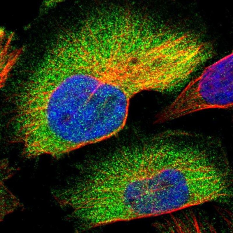
- Main image

- Experimental details
- Immunofluorescent staining of human cell line U-251 MG shows localization to cytosol.
- Sample type
- Human
Supportive validation
- Submitted by
- Atlas Antibodies (provider)
- Enhanced method
- Orthogonal validation
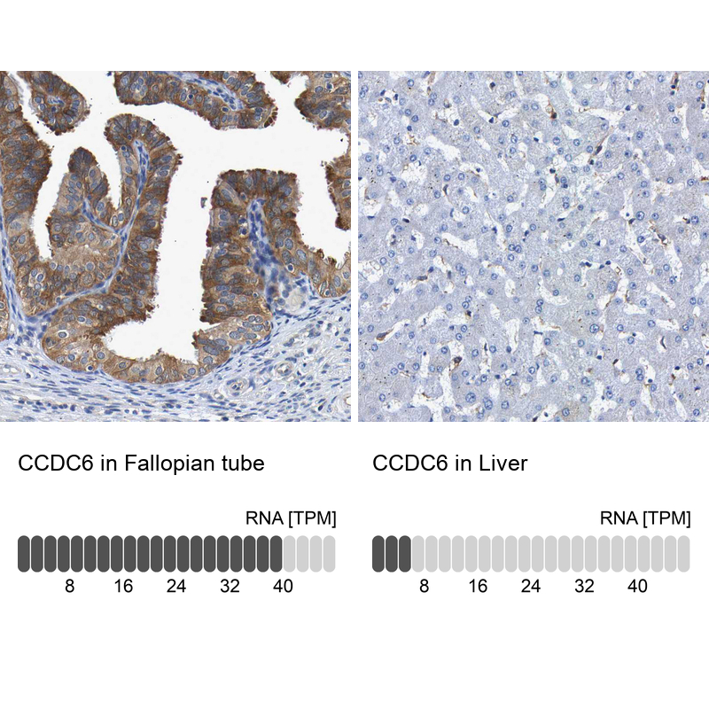
- Main image

- Experimental details
- Immunohistochemistry analysis in human fallopian tube and liver tissues using HPA019051 antibody. Corresponding CCDC6 RNA-seq data are presented for the same tissues.
- Sample type
- Human
- Protocol
- Protocol