Explore
 Validate
 Learn
Learn Western blot
Antibody data
- Antibody Data
- Antigen structure
- References [0]
- Comments [0]
- Validations
- Western blot [2]
- Immunohistochemistry [3]
Submit
Validation data
Reference
Comment
Report error
- Product number
- MA1-100 - Provider product page

- Provider
- Invitrogen Antibodies
- Product name
- p38 MAPK gamma Monoclonal Antibody (10E1)
- Antibody type
- Monoclonal
- Antigen
- Other
- Description
- MA1-100 has been successfully used in Western blot and immunohistochemistry applications and reacts with human and mouse samples. MA1-100 specifically recognizes p38-gamma, and does not react with alpha, beta, or delta isoforms.
- Reactivity
- Human, Mouse, Canine
- Host
- Mouse
- Isotype
- IgG
- Antibody clone number
- 10E1
- Vial size
- 100 μg
- Concentration
- 1 mg/mL
- Storage
- -20°C
No comments: Submit comment
Supportive validation
- Submitted by
- Invitrogen Antibodies (provider)
- Main image

- Experimental details
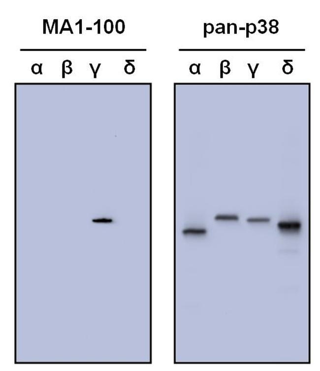
- Western blot analysis of p38 was performed by loading 50 ng of human, recombinant p38-alpha, beta, gamma and delta per well onto a Novex 4-20% Tris-Glycine polyacrylamide gel (Product # WT4202BOX ). Proteins were transferred to a nitrocellulose membrane using the G2 Blotter (Product # 62288), and blocked with 5% milk in TBST for one hour at room temperature. Isoforms of p38 were detected using a p38 monoclonal antibody (Product # MA1-100, left panel) and a pan-p38 antibody (right panel) at a dilution of 1 µg/mL in blocking buffer overnight at 4C on a rocking platform, followed by a Goat anti-mouse IgG HRP-linked secondary antibody (Product # 31430) at a dilution of 1:10,000 for at least 30 minutes. Chemiluminescent detection was performed using SuperSignal Pico (Product # 34078) and the myECL™ Imager (Product # 62236)
- Submitted by
- Invitrogen Antibodies (provider)
- Main image

- Experimental details
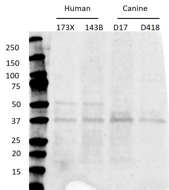
- Western blot analysis of p38 MAPK gamma was performed by loading whole-cell lysates of human osteosarcoma cell lines 143B and 17-3X and canine cell lines D17 and D418. p38 MAPK gamma was detected at approximately 38 kDa using a p38 MAPK alpha monoclonal antibody (Product # MA1-100) at a dilution of 1:1000 in Blocking Buffer (Product # 37543) overnight at 4°C on a rocking platform, followed by a 1-hour room-temperature incubation of a goat anti-mouse IgG antibody at a dilution of 1:5000. Data courtesy of Thermo Scientific KOL Program.
Supportive validation
- Submitted by
- Invitrogen Antibodies (provider)
- Main image
- Experimental details
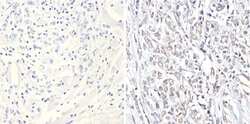
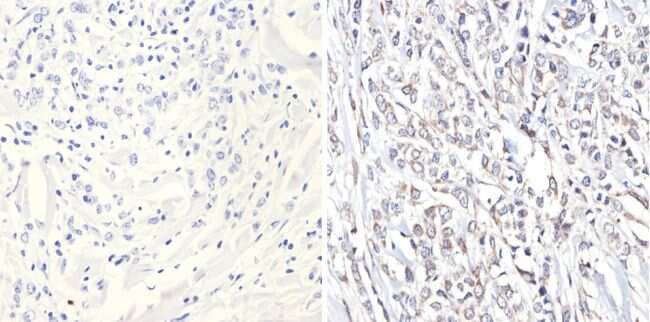
- Immunohistochemistry analysis of MAPK12 showing positive staining in the nucleus and cytoplasm of paraffin-treated Human breast carcinoma (right) compared with a negative control in the absence of primary antibody (left). To expose target proteins, antigen retrieval method was performed using 10mM sodium citrate (pH 6.0), microwaved for 8-15 min. Following antigen retrieval, tissues were blocked in 3% H2O2-methanol for 15 min at room temperature, washed with ddH2O and PBS, and then probed with a MAPK12 monoclonal antibody (Product # MA1-100) diluted by 3% BSA-PBS at a dilution of 1:20 overnight at 4°C in a humidified chamber. Tissues were washed extensively PBST and detection was performed using an HRP-conjugated secondary antibody followed by colorimetric detection using a DAB kit. Tissues were counterstained with hematoxylin and dehydrated with ethanol and xylene to prep for mounting.
- Submitted by
- Invitrogen Antibodies (provider)
- Main image

- Experimental details
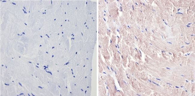
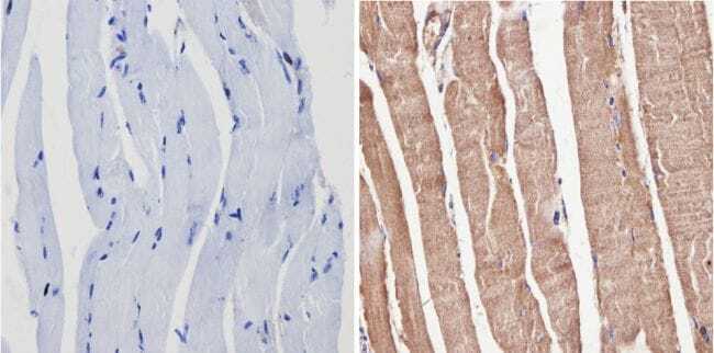
- Immunohistochemistry analysis of MAPK12 showing positive staining in the nucleus and cytoplasm of paraffin-treated Mouse skeletal muscle (right) compared with a negative control in the absence of primary antibody (left). To expose target proteins, antigen retrieval method was performed using 10mM sodium citrate (pH 6.0), microwaved for 8-15 min. Following antigen retrieval, tissues were blocked in 3% H2O2-methanol for 15 min at room temperature, washed with ddH2O and PBS, and then probed with a MAPK12 monoclonal antibody (Product # MA1-100) diluted by 3% BSA-PBS at a dilution of 1:20 overnight at 4°C in a humidified chamber. Tissues were washed extensively PBST and detection was performed using an HRP-conjugated secondary antibody followed by colorimetric detection using a DAB kit. Tissues were counterstained with hematoxylin and dehydrated with ethanol and xylene to prep for mounting.
- Submitted by
- Invitrogen Antibodies (provider)
- Main image

- Experimental details
- Immunohistochemistry analysis of MAPK12 showing positive staining in the nucleus and cytoplasm of paraffin-treated Human skeletal muscle (right) compared with a negative control in the absence of primary antibody (left). To expose target proteins, antigen retrieval method was performed using 10mM sodium citrate (pH 6.0), microwaved for 8-15 min. Following antigen retrieval, tissues were blocked in 3% H2O2-methanol for 15 min at room temperature, washed with ddH2O and PBS, and then probed with a MAPK12 monoclonal antibody (Product # MA1-100) diluted by 3% BSA-PBS at a dilution of 1:20 overnight at 4°C in a humidified chamber. Tissues were washed extensively PBST and detection was performed using an HRP-conjugated secondary antibody followed by colorimetric detection using a DAB kit. Tissues were counterstained with hematoxylin and dehydrated with ethanol and xylene to prep for mounting.