Explore
 Validate
 Learn
Learn Western blot
Antibody data
- Antibody Data
- Antigen structure
- References [1]
- Comments [0]
- Validations
- Western blot [2]
- Immunocytochemistry [1]
Submit
Validation data
Reference
Comment
Report error
- Product number
- MAB1347 - Provider product page

- Provider
- R&D Systems
- Product name
- Human/Mouse/Rat p38 gamma Antibody
- Antibody type
- Monoclonal
- Description
- Protein A or G purified from hybridoma culture supernatant. Detects human, mouse, and rat p38 gamma in direct ELISAs and Western blots. In Western blots, no cross-reactivity with recombinant p38 alpha , p38 beta , or p38 delta is observed.
- Reactivity
- Human, Mouse, Rat
- Host
- Mouse
- Conjugate
- Unconjugated
- Antigen sequence
P53778- Isotype
- IgG
- Antibody clone number
- 212464
- Vial size
- 100 ug
- Concentration
- LYOPH
- Storage
- Use a manual defrost freezer and avoid repeated freeze-thaw cycles. 12 months from date of receipt, -20 to -70 °C as supplied. 1 month, 2 to 8 °C under sterile conditions after reconstitution. 6 months, -20 to -70 °C under sterile conditions after reconstitution.
Submitted references Differential tissue expression and activation of p38 MAPK alpha, beta, gamma, and delta isoforms in rheumatoid arthritis.
Korb A, Tohidast-Akrad M, Cetin E, Axmann R, Smolen J, Schett G
Arthritis and rheumatism 2006 Sep;54(9):2745-56
Arthritis and rheumatism 2006 Sep;54(9):2745-56
No comments: Submit comment
Supportive validation
- Submitted by
- R&D Systems (provider)
- Main image
- Experimental details
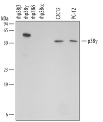
- Detection of Human/Mouse/Rat p38 gamma by Western Blot. Western blot shows lysates of C2C12 mouse myoblast cell line myoblast cell line and PC-12 rat adrenal pheochromocytoma cell line. PVDF membrane was probed with 1 µg/mL Mouse Anti-Human/Mouse/Rat p38 gamma Monoclonal Antibody (Catalog # MAB1347) followed by HRP-conjugated Anti-Mouse IgG Secondary Antibody (Catalog # HAF007). For additional reference, recombinant human p38 beta , p38 gamma , p38 delta and p38 alpha (2 ng/lane) were included. A specific band for native p38 gamma was detected at approximately 40 kDa (as indicated). This experiment was conducted under reducing conditions and using Immunoblot Buffer Group 4.

- Submitted by
- R&D Systems (provider)
- Main image
- Experimental details
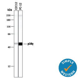
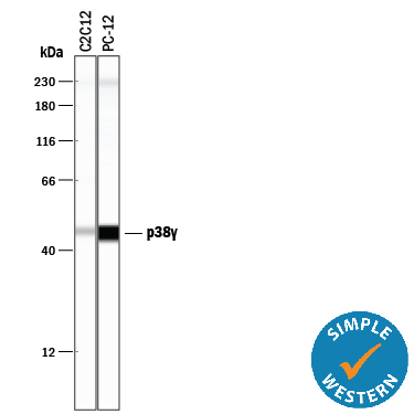
- Detection of Mouse and Rat p38 gamma by Simple WesternTM. Simple Western lane view shows lysates of C2C12 mouse myoblast cell line and PC-12 rat adrenal pheochromocytoma cell line, loaded at 0.5 mg/mL. A specific band was detected for p38 gamma at approximately 46 kDa (as indicated) using 10 µg/mL of Mouse Anti-Human/Mouse/Rat p38 gamma Monoclonal Antibody (Catalog # MAB1347). This experiment was conducted under reducing conditions and using the 12-230 kDa separation system.
Supportive validation
- Submitted by
- R&D Systems (provider)
- Main image
- Experimental details
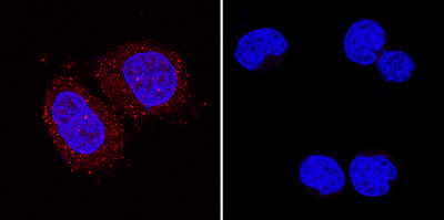
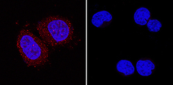
- p38 gamma in MCF-7 and MOLT-4 Human Cell Lines. p38 gamma was detected in immersion fixed MCF-7 human breast cancer cell line (positive control, left panel) and MOLT-4 human acute lymphoblastic leukemia cell line (negative control, right panel) using Mouse Anti-Human/Mouse/Rat p38 gamma Monoclonal Antibody (Catalog # MAB1347) at 5 µg/mL for 3 hours at room temperature. Cells were stained using the NorthernLights™ 557-conjugated Anti-Mouse IgG Secondary Antibody (red; Catalog # NL007) and counterstained with DAPI (blue). Specific staining was localized to cytoplasm in MCF-7 cells. View our protocol for Fluorescent ICC Staining of Cells on Coverslips.