Explore
 Validate
 Learn
Learn Western blot
 ELISA
ELISA Immunocytochemistry
Antibody data
- Antibody Data
- Antigen structure
- References [0]
- Comments [0]
- Validations
- Immunocytochemistry [6]
- Immunoprecipitation [1]
- Immunohistochemistry [2]
Submit
Validation data
Reference
Comment
Report error
- Product number
- MA5-35115 - Provider product page

- Provider
- Invitrogen Antibodies
- Product name
- MCM3 Recombinant Rabbit Monoclonal Antibody (9Z5W4)
- Antibody type
- Monoclonal
- Antigen
- Synthetic peptide
- Description
- Immunogen sequence: MAGTVVLDDV ELREAQRDYL DFLDDEEDQG IYQSKVRELI SDNQYRLIVN VNDLRRKNEK RANRLLNNAF EELVAFQRAL KDFVASIDAT YAKQYEEFYV
- Reactivity
- Human, Mouse, Rat
- Host
- Rabbit
- Isotype
- IgG
- Antibody clone number
- 9Z5W4
- Vial size
- 100 μL
- Concentration
- 0.4 mg/mL
- Storage
- -20°C, Avoid Freeze/Thaw Cycles
No comments: Submit comment
Supportive validation
- Submitted by
- Invitrogen Antibodies (provider)
- Main image
- Experimental details
- Immunocytochemical analysis of MCM3 performed on HeLa cells using a MCM3 monoclonal antibody (Product # MA5-35115) at a dilution of 1:100. An HRP Goat Anti-Rabbit IgG (H+L) secondary antibody was applied at a dilution of 1:10,000.

- Submitted by
- Invitrogen Antibodies (provider)
- Main image
- Experimental details
- Immunocytochemical analysis of MCM3 performed on NIH-3T3 cells using a MCM3 monoclonal antibody (Product # MA5-35115) at a dilution of 1:100. An HRP Goat Anti-Rabbit IgG (H+L) secondary antibody was applied at a dilution of 1:10,000.
- Submitted by
- Invitrogen Antibodies (provider)
- Main image

- Experimental details
- Immunocytochemical analysis of MCM3 performed on HeLa cells using a MCM3 monoclonal antibody (Product # MA5-35115) at a dilution of 1:100. An HRP Goat Anti-Rabbit IgG (H+L) secondary antibody was applied at a dilution of 1:10,000.
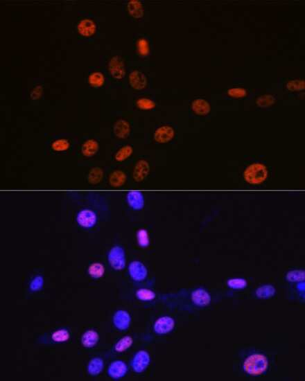
- Submitted by
- Invitrogen Antibodies (provider)
- Main image

- Experimental details
- Immunocytochemical analysis of MCM3 performed on NIH-3T3 cells using a MCM3 monoclonal antibody (Product # MA5-35115) at a dilution of 1:100. An HRP Goat Anti-Rabbit IgG (H+L) secondary antibody was applied at a dilution of 1:10,000.
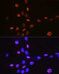
- Submitted by
- Invitrogen Antibodies (provider)
- Main image

- Experimental details
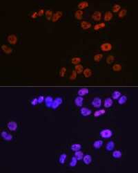
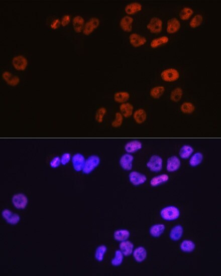
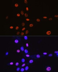
- Immunofluorescence analysis of MCM3 in HeLa cells. Samples were incubated with MCM3 Monoclonal antibody (Product # MA5-35115) using a dilution of 1:100 (40x lens). Blue: DAPI for nuclear staining.

- Submitted by
- Invitrogen Antibodies (provider)
- Main image
- Experimental details
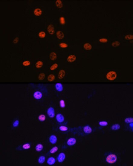
- Immunofluorescence analysis of MCM3 in NIH-3T3 cells. Samples were incubated with MCM3 Monoclonal antibody (Product # MA5-35115) using a dilution of 1:100 (40x lens). Blue: DAPI for nuclear staining.

Supportive validation
- Submitted by
- Invitrogen Antibodies (provider)
- Main image

- Experimental details
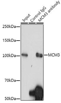
- Immunoprecipitation of MCM3 in 300 μg extracts of Hela cells. Samples were precipitated with 3 μg MCM3 Monoclonal antibody (Product # MA5-35115). Western blot was performed from the immunoprecipitate using MCM3 Monoclonal antibody (Product # MA5-35115) at a dilution of 1:1,000.
Supportive validation
- Submitted by
- Invitrogen Antibodies (provider)
- Main image
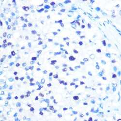
- Experimental details
- Immunohistochemistry analysis of MCM3 in paraffin-embedded human liver cancer. Samples were incubated with MCM3 Monoclonal antibody (Product # MA5-35115) using a dilution of 1:100 (40x lens). Perform microwave antigen retrieval with 10 mM PBS buffer pH 7.2 before commencing with IHC staining protocol.
- Submitted by
- Invitrogen Antibodies (provider)
- Main image

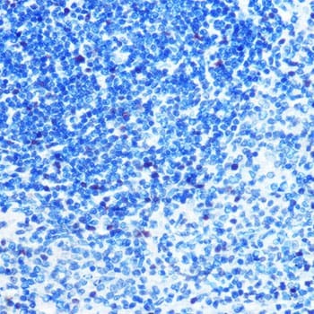
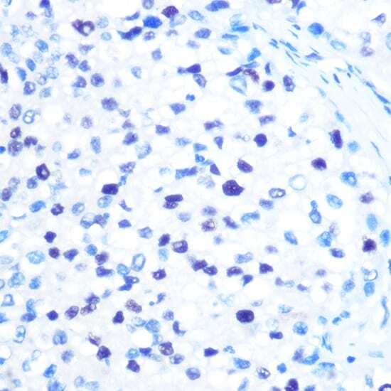
- Experimental details
- Immunohistochemistry analysis of MCM3 in paraffin-embedded rat spleen. Samples were incubated with MCM3 Monoclonal antibody (Product # MA5-35115) using a dilution of 1:100 (40x lens). Perform microwave antigen retrieval with 10 mM PBS buffer pH 7.2 before commencing with IHC staining protocol.