Explore
 Validate
 Learn
Learn Western blot
 Immunocytochemistry
Antibody data
- Antibody Data
- Antigen structure
- References [3]
- Comments [0]
- Validations
- Immunocytochemistry [1]
- Blocking/Neutralizing [1]
Submit
Validation data
Reference
Comment
Report error
- Product number
- MAB203-100 - Provider product page

- Provider
- R&D Systems
- Product name
- Human IL-3 Antibody
- Antibody type
- Monoclonal
- Description
- Protein A or G purified from ascites. Detects human IL-3 in direct ELISAs and Western blots. In direct ELISAs and Western blots, no cross-reactivity with recombinant mouse IL-3 is observed. Does not neutralize the biological activity of recombinant mouse IL-3.
- Reactivity
- Human
- Host
- Mouse
- Conjugate
- Unconjugated
- Antigen sequence
Q6GS87- Isotype
- IgG
- Antibody clone number
- 4806
- Vial size
- 100 ug
- Concentration
- LYOPH
- Storage
- Use a manual defrost freezer and avoid repeated freeze-thaw cycles. 12 months from date of receipt, -20 to -70 °C as supplied. 1 month, 2 to 8 °C under sterile conditions after reconstitution. 6 months, -20 to -70 °C under sterile conditions after reconstitution.
Submitted references Hematopoietic stem cell cytokines and fibroblast growth factor-2 stimulate human endothelial cell-pericyte tube co-assembly in 3D fibrin matrices under serum-free defined conditions.
Human T-cell leukemia virus type 2 induces survival and proliferation of CD34(+) TF-1 cells through activation of STAT1 and STAT5 by secretion of interferon-gamma and granulocyte macrophage-colony-stimulating factor.
Expression of functional interleukin-3 receptors on Hodgkin and Reed-Sternberg cells.
Smith AO, Bowers SL, Stratman AN, Davis GE
PloS one 2013;8(12):e85147
PloS one 2013;8(12):e85147
Human T-cell leukemia virus type 2 induces survival and proliferation of CD34(+) TF-1 cells through activation of STAT1 and STAT5 by secretion of interferon-gamma and granulocyte macrophage-colony-stimulating factor.
Bovolenta C, Pilotti E, Mauri M, Turci M, Ciancianaini P, Fisicaro P, Bertazzoni U, Poli G, Casoli C
Blood 2002 Jan 1;99(1):224-31
Blood 2002 Jan 1;99(1):224-31
Expression of functional interleukin-3 receptors on Hodgkin and Reed-Sternberg cells.
Aldinucci D, Poletto D, Gloghini A, Nanni P, Degan M, Perin T, Ceolin P, Rossi FM, Gattei V, Carbone A, Pinto A
The American journal of pathology 2002 Feb;160(2):585-96
The American journal of pathology 2002 Feb;160(2):585-96
No comments: Submit comment
Supportive validation
- Submitted by
- R&D Systems (provider)
- Main image
- Experimental details
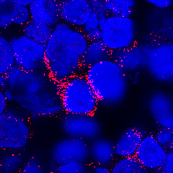
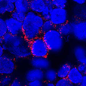
- IL-3 in Human PBMCs. IL-3 was detected in immersion fixed human peripheral blood mononuclear cells (PBMCs) using Mouse Anti-Human IL-3 Monoclonal Antibody (Catalog # MAB203) at 25 µg/mL for 3 hours at room temperature. Cells were stained using the NorthernLights™ 557-conjugated Anti-Mouse IgG Secondary Antibody (red; Catalog # NL007) and counterstained with DAPI (blue). Specific staining was localized to cytoplasm. View our protocol for Fluorescent ICC Staining of Cells on Coverslips.
Supportive validation
- Submitted by
- R&D Systems (provider)
- Main image
- Experimental details
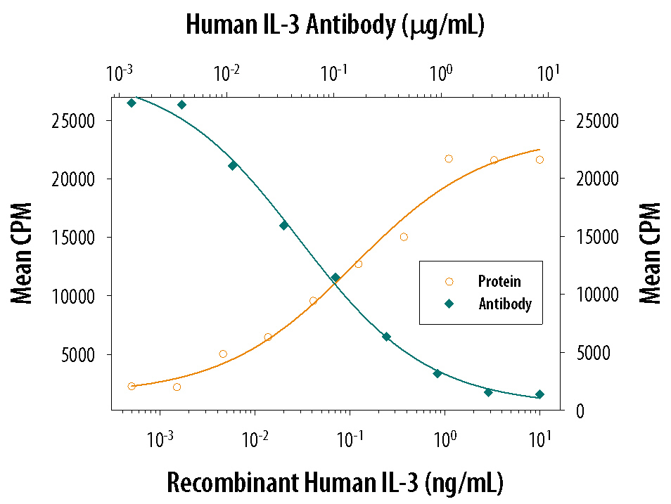
- Cell Proliferation Induced by IL-3 and Neutralization by Human IL-3 Antibody. Recombinant Human IL-3 (Catalog # 203-IL) stimulates proliferation in the TF-1 human erythroleukemic cell line in a dose-dependent manner (orange line). Proliferation elicited by Recombinant Human IL-3 (1.25 ng/mL) is neutralized (green line) by increasing concentrations of Mouse Anti-Human IL-3 Monoclonal Antibody (Catalog # MAB203). The ND50 is typically 0.03-0.08 µg/mL.