Explore
 Validate
 Learn
Learn Western blot
Antibody data
- Antibody Data
- Antigen structure
- References [3]
- Comments [0]
- Validations
- Western blot [1]
Submit
Validation data
Reference
Comment
Report error
- Product number
- PAB16927 - Provider product page

- Provider
- Abnova Corporation
- Proper citation
- Abnova Corporation Cat#PAB16927, RRID:AB_10677226
- Product name
- Csf2 polyclonal antibody
- Antibody type
- Polyclonal
- Description
- Rabbit polyclonal antibody raised against full length recombinant Csf2.
- Storage
- Store at 4°C on dry atmosphere.After reconstitution with 0.1 mL of deionized water, store at -20°C or lower.Aliquot to avoid repeated freezing and thawing.
Submitted references Recombinant murine GM-CSF from E. coli has biological activity and is neutralized by a specific antiserum.
The structure and expression of the murine gene encoding granulocyte-macrophage colony stimulating factor: evidence for utilisation of alternative promoters.
Structure of the chromosomal gene for granulocyte-macrophage colony stimulating factor: comparison of the mouse and human genes.
DeLamarter JF, Mermod JJ, Liang CM, Eliason JF, Thatcher DR
The EMBO journal 1985 Oct;4(10):2575-81
The EMBO journal 1985 Oct;4(10):2575-81
The structure and expression of the murine gene encoding granulocyte-macrophage colony stimulating factor: evidence for utilisation of alternative promoters.
Stanley E, Metcalf D, Sobieszczuk P, Gough NM, Dunn AR
The EMBO journal 1985 Oct;4(10):2569-73
The EMBO journal 1985 Oct;4(10):2569-73
Structure of the chromosomal gene for granulocyte-macrophage colony stimulating factor: comparison of the mouse and human genes.
Miyatake S, Otsuka T, Yokota T, Lee F, Arai K
The EMBO journal 1985 Oct;4(10):2561-8
The EMBO journal 1985 Oct;4(10):2561-8
No comments: Submit comment
Supportive validation
- Submitted by
- Abnova Corporation (provider)
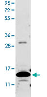
- Main image

- Experimental details
- Western blot using Csf2 polyclonal antibody (Cat # PAB16927) shows detection of a band ~14 KDa in size corresponding to recombinant mouse Csf2. After transfer, the membrane was blocked overnight with 3% BSA in TBS followed by reaction with primary antibody at a 1 : 1,000 dilution. Detection occurred using DyLight™649 conjugated anti-Rabbit IgG secondary antibody diluted 1 : 20,000 in blocking buffer. Image was captured using VersaDoc MP 4000 imaging system (Bio-Rad).