Explore
 Validate
 Learn
Learn Western blot
 Immunohistochemistry
Antibody data
- Antibody Data
- Antigen structure
- References [4]
- Comments [0]
- Validations
- Immunohistochemistry [1]
Submit
Validation data
Reference
Comment
Report error
- Product number
- HPA018849 - Provider product page

- Provider
- Atlas Antibodies
- Proper citation
- Atlas Antibodies Cat#HPA018849, RRID:AB_1848310
- Product name
- Anti-EVL
- Antibody type
- Polyclonal
- Description
- Polyclonal Antibody against Human EVL, Gene description: Enah/Vasp-like, Alternative Gene Names: RNB6, Validated applications: IHC, WB, Uniprot ID: Q9UI08, Storage: Store at +4°C for short term storage. Long time storage is recommended at -20°C.
- Reactivity
- Human
- Host
- Rabbit
- Conjugate
- Unconjugated
- Isotype
- IgG
- Vial size
- 100 µl
- Concentration
- 0.05 mg/ml
- Storage
- Store at +4°C for short term storage. Long time storage is recommended at -20°C.
- Handling
- The antibody solution should be gently mixed before use.
Submitted references A distributed residue network permits conformational binding specificity in a conserved family of actin remodelers
Mechanosensing during directed cell migration requires dynamic actin polymerization at focal adhesions
The actin cytoskeletal architecture of estrogen receptor positive breast cancer cells suppresses invasion
Actin stress fiber organization promotes cell stiffening and proliferation of pre-invasive breast cancer cells
Hwang T, Parker S, Hill S, Ilunga M, Grant R, Mouneimne G, Keating A
eLife 2021;10
eLife 2021;10
Mechanosensing during directed cell migration requires dynamic actin polymerization at focal adhesions
Puleo J, Parker S, Roman M, Watson A, Eliato K, Peng L, Saboda K, Roe D, Ros R, Gertler F, Mouneimne G
Journal of Cell Biology 2019;218(12):4215-4235
Journal of Cell Biology 2019;218(12):4215-4235
The actin cytoskeletal architecture of estrogen receptor positive breast cancer cells suppresses invasion
Padilla-Rodriguez M, Parker S, Adams D, Westerling T, Puleo J, Watson A, Hill S, Noon M, Gaudin R, Aaron J, Tong D, Roe D, Knudsen B, Mouneimne G
Nature Communications 2018;9(1)
Nature Communications 2018;9(1)
Actin stress fiber organization promotes cell stiffening and proliferation of pre-invasive breast cancer cells
Tavares S, Vieira A, Taubenberger A, Araújo M, Martins N, Brás-Pereira C, Polónia A, Herbig M, Barreto C, Otto O, Cardoso J, Pereira-Leal J, Guck J, Paredes J, Janody F
Nature Communications 2017;8(1)
Nature Communications 2017;8(1)
No comments: Submit comment
Supportive validation
- Submitted by
- Atlas Antibodies (provider)
- Enhanced method
- Orthogonal validation
- Main image
- Experimental details
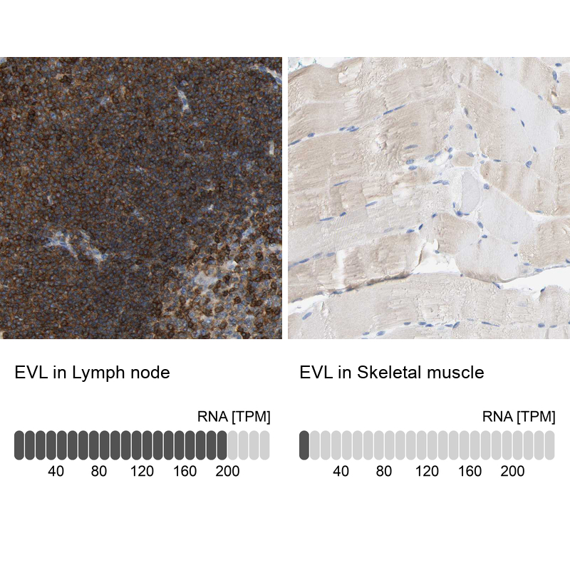
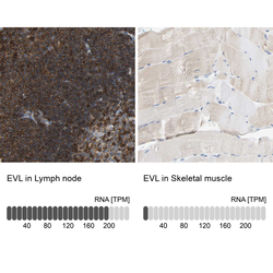
- Immunohistochemistry analysis in human lymph node and skeletal muscle tissues using HPA018849 antibody. Corresponding EVL RNA-seq data are presented for the same tissues.
- Sample type
- Human
- Protocol
- Protocol