 Explore
 Validate
 Learn
Learn Western blot
 Flow cytometry
Antibody data
- Antibody Data
- Antigen structure
- References [0]
- Comments [0]
- Validations
- Western blot [4]
Submit
Validation data
Reference
Comment
Report error
- Product number
- MA5-25393 - Provider product page

- Provider
- Invitrogen Antibodies
- Product name
- Gephyrin Monoclonal Antibody (OTI3A6)
- Antibody type
- Monoclonal
- Antigen
- Recombinant full-length protein
- Reactivity
- Human, Mouse, Rat
- Host
- Mouse
- Isotype
- IgG
- Antibody clone number
- OTI3A6
- Vial size
- 100 µL
- Concentration
- 1 mg/mL
- Storage
- -20° C, Avoid Freeze/Thaw Cycles
No comments: Submit comment
Supportive validation
- Submitted by
- Invitrogen Antibodies (provider)
- Main image
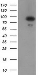
- Experimental details
- Western blot analysis of GPHN in HEK293T cells in untransfected (Left lane) and transfected (Right lane) samples using 5 µg per lane. The samples were separated by SDS-PAGE and probed with GPHN (Product # MA5-25393) monoclonal antibody.

- Submitted by
- Invitrogen Antibodies (provider)
- Main image

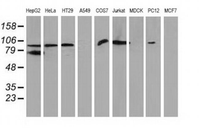
- Experimental details
- Western blot analysis of GPHN in HepG2, HeLa, SVT2, A549, COS7, Jurkat, MDCK, PC12 and MCF7 cells using 35 µg per lane. Samples were separated by SDS-PAGE and probed with GPHN (Product # MA5-25393) monoclonal antibody.
- Submitted by
- Invitrogen Antibodies (provider)
- Main image

- Experimental details
- Knockdown of GPHN was achieved by transfecting PC-3 with GPHN specific siRNAs (Silencer® select Product # s20000, s20001). Western blot analysis (Fig. a) was performed using whole cell extracts from GPHN knockdown cells (Lane 3), non-specific scrambled siRNA transfected cells (Lane 2) and untransfected cells (Lane 1). The blot was probed with Gephyrin Monoclonal Antibody (OTI3A6) (Product # MA5-25393, 1:1200 dilution) and Goat anti-Mouse IgG (H+L) Superclonal™ Recombinant Secondary Antibody, HRP (Product # A28177, 1:4000 dilution). Densitometric analysis of this western blot is shown in histogram (Fig. b). Decrease in signal upon siRNA mediated knock down confirms that antibody is specific to GPHN.

- Submitted by
- Invitrogen Antibodies (provider)
- Main image

- Experimental details
- Western blot was performed using Anti-Gephyrin Monoclonal Antibody (OTI3A6) (Product # MA5-25393) and 93 kDa, 150 kDa and 250 kDa bands corresponding to GPHN was observed across all the cell lines and tissues tested. Whole cell extracts (30 µg lysate) of IMR32 (Lane 1), Neuro-2a (Lane 2), PC-3 (Lane 3), NIH/3T3 (Lane 4), PC-12 (Lane 5) and tissue extracts of Mouse Brain (Lane 6) and Rat Brain (Lane 7) were electrophoresed using NuPAGE™ 4-12% Bis-Tris Protein Gel (Product # NP0322BOX). Resolved proteins were then transferred onto a nitrocellulose membrane (Product # IB23001) by iBlot® 2 Dry Blotting System (Product # IB21001). The blot was probed with the primary antibody (1:1200 dilution) and detected by chemiluminescence with Goat anti-Mouse IgG (H+L) Superclonal™ Recombinant Secondary Antibody, HRP (Product # A28177, 1:4000 dilution) using the iBright FL 1000 (Product # A32752). Chemiluminescent detection was performed using Novex® ECL Chemiluminescent Substrate Reagent Kit (Product # WP20005). The 150 kDa and 250 kDa bands observed in PC-12 are reported to be SUMOylated form of GPHN. [doi: 10.1038/ncomms13365 (2016)] and the 25 kDa band in Mouse Brain is the circulating IgGs that are commonly detected in tissues..
