Explore
 Validate
 Learn
Learn Western blot
Antibody data
- Antibody Data
- Antigen structure
- References [0]
- Comments [0]
- Validations
- Western blot [5]
- Immunocytochemistry [1]
- Immunoprecipitation [1]
- Immunohistochemistry [2]
Submit
Validation data
Reference
Comment
Report error
- Product number
- GTX109734 - Provider product page

- Provider
- GeneTex
- Proper citation
- GeneTex Cat#GTX109734, RRID:AB_11164321
- Product name
- Gephyrin antibody [N2C1], Internal
- Antibody type
- Polyclonal
- Reactivity
- Human, Mouse, Rat
- Host
- Rabbit
No comments: Submit comment
Supportive validation
- Submitted by
- GeneTex (provider)
- Main image
- Experimental details
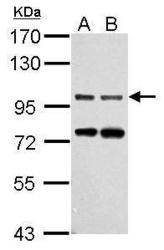
- Sample (30 ug of whole cell lysate) A: NIH-3T3 B: JC 7.5% SDS PAGE GTX109734 diluted at 1:1000
- Submitted by
- GeneTex (provider)
- Main image

- Experimental details
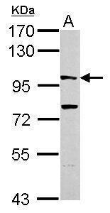
- Sample (30 ug of whole cell lysate) A: PC-12 7.5% SDS PAGE GTX109734 diluted at 1:1000
- Submitted by
- GeneTex (provider)
- Main image
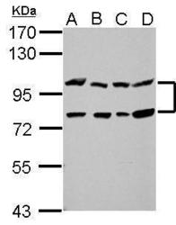
- Experimental details
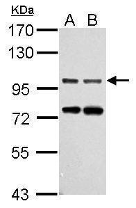
- Sample (30 ug of whole cell lysate) A: 293T B: A431 C: HeLa D: HepG2 7.5% SDS PAGE GTX109734 diluted at 1:1000
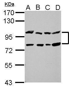
- Submitted by
- GeneTex (provider)
- Main image
- Experimental details
- Various whole cell extracts (30 ?g) were separated by 7.5% SDS-PAGE, and the membrane was blotted with Gephyrin antibody [N2C1], Internal (GTX109734) diluted at 1:3000. The HRP-conjugated anti-rabbit IgG antibody (GTX213110-01) was used to detect the primary antibody.
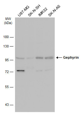
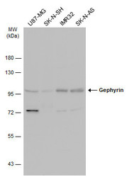
- Submitted by
- GeneTex (provider)
- Main image
- Experimental details
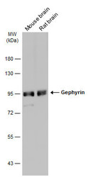
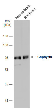
- Various tissue extracts (50 ?g) were separated by 7.5% SDS-PAGE, and the membrane was blotted with Gephyrin antibody [N2C1], Internal (GTX109734) diluted at 1:5000. The HRP-conjugated anti-rabbit IgG antibody (GTX213110-01) was used to detect the primary antibody.
Supportive validation
- Submitted by
- GeneTex (provider)
- Main image
- Experimental details
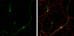
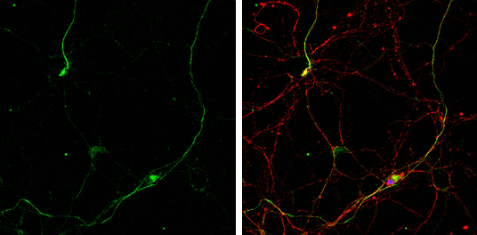
- Gephyrin antibody [N2C1], Internal detects Gephyrin protein by immunofluorescent analysis.Sample: DIV10 rat E18 primary hippocampal neurons were fixed in 4% paraformaldehyde at RT for 15 min.Green: Gephyrin protein stained by Gephyrin antibody [N2C1], Internal (GTX109734) diluted at 1:500.Red: beta Tubulin 3/ Tuj1, stained by beta Tubulin 3/ Tuj1 antibody [GT1338] (GTX631831) diluted at 1:500.Blue: Fluoroshield with DAPI (GTX30920).
Supportive validation
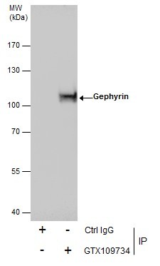
- Submitted by
- GeneTex (provider)
- Main image
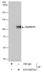
- Experimental details
- Immunoprecipitation of Gephyrin protein from A431 whole cell extracts using 5 £gg of Gephyrin antibody [N2C1], Internal (GTX109734).Western blot analysis was performed using Gephyrin antibody [N2C1], Internal (GTX109734).EasyBlot anti-Rabbit IgG (GTX221666-01) was used as a secondary reagent.
Supportive validation
- Submitted by
- GeneTex (provider)
- Main image

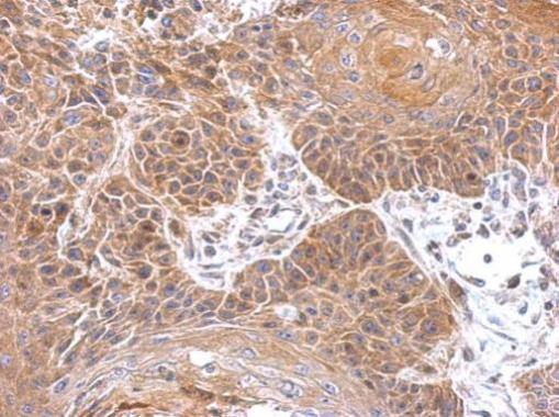
- Experimental details
- Immunohistochemical analysis of paraffin-embedded mouse brain, using Gephyrin(GTX109734) antibody at 1:500 dilution.
- Submitted by
- GeneTex (provider)
- Main image
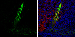
- Experimental details
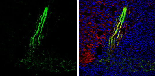
- Gephyrin antibody [N2C1], Internal detects Gephyrin protein expression by immunohistochemical analysis.Sample: Frozen sectioned E13.5 Rat brain. Green: Gephyrin protein stained by Gephyrin antibody [N2C1], Internal (GTX109734) diluted at 1:250.Red: beta Tubulin 3/ TUJ1, a mature neuron marker, stained by beta Tubulin 3/ TUJ1 antibody [GT11710] (GTX631836) diluted at 1:500.Blue: Fluoroshield with DAPI (GTX30920).