 Explore
 Validate
 Learn
Learn Western blot
 Immunocytochemistry
Antibody data
- Antibody Data
- Antigen structure
- References [1]
- Comments [0]
- Validations
- Immunocytochemistry [4]
- Immunoprecipitation [1]
- Immunohistochemistry [2]
- Other assay [1]
Submit
Validation data
Reference
Comment
Report error
- Product number
- PA5-29036 - Provider product page

- Provider
- Invitrogen Antibodies
- Product name
- Gephyrin Polyclonal Antibody
- Antibody type
- Polyclonal
- Antigen
- Recombinant full-length protein
- Description
- Recommended positive controls: NIH-3T3, JC, U87-MG, SK-N-SH, IMR-32, SK-N-AS, mouse brain, rat brain. Predicted reactivity: Mouse (100%), Rat (100%), Xenopus laevis (92%), Pig (100%), Chicken (100%), Rhesus Monkey (100%). Store product as a concentrated solution. Centrifuge briefly prior to opening the vial.
- Reactivity
- Human, Mouse, Rat
- Host
- Rabbit
- Isotype
- IgG
- Vial size
- 100 μL
- Concentration
- 0.59 mg/mL
- Storage
- Store at 4°C short term. For long term storage, store at -20°C, avoiding freeze/thaw cycles.
Submitted references Immunohistochemical Characterization of the Nervous System of Culex pipiens (Diptera, Culicidae).
Gregor KM, Becker SC, Hellhammer F, Baumgärtner W, Puff C
Biology 2022 Jan 1;11(1)
Biology 2022 Jan 1;11(1)
No comments: Submit comment
Supportive validation
- Submitted by
- Invitrogen Antibodies (provider)
- Main image

- Experimental details
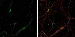
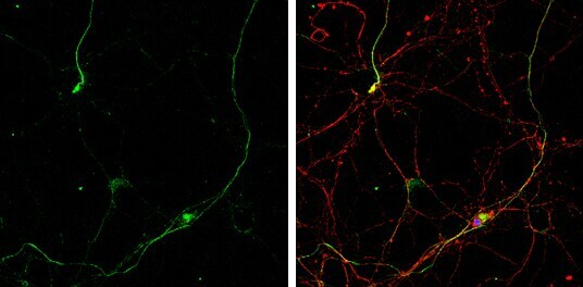
- Immunocytochemistry-Immunofluorescence analysis of Gephyrin was performed in DIV10 rat E18 primary hippocampal neurons fixed in 4% paraformaldehyde at RT for 15 min. Green: Gephyrin Polyclonal Antibody (Product # PA5-29036) diluted at 1:500. Red: beta Tubulin 3/ Tuj1, stained by beta Tubulin 3/ Tuj1 antibody. Blue: Fluoroshield with DAPI.

- Submitted by
- Invitrogen Antibodies (provider)
- Main image
- Experimental details
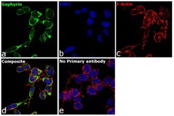
- Immunofluorescence analysis of Gephyrin was performed using 70% confluent log phase Neuro-2a cells. The cells were fixed with 4% paraformaldehyde for 10 minutes, permeabilized with 0.1% Triton™ X-100 for 15 minutes, and blocked with 1% BSA for 1 hour at room temperature. The cells were labeled with Gephyrin Rabbit Polyclonal Antibody (Product # PA5-29036) at 5 microgram/mL in 0.1% BSA, incubated at 4 degree Celsius overnight and then labeled with Goat anti-Rabbit IgG (H+L) Superclonal™ Secondary Antibody, Alexa Fluor® 488 conjugate (Product # A27034) at a dilution of 1:2000 for 45 minutes at room temperature (Panel a: green). Nuclei (Panel b: blue) were stained with ProLong™ Diamond Antifade Mountant with DAPI (Product # P36962). F-actin (Panel c: red) was stained with Rhodamine Phalloidin (Product # R415). Panel d represents the merged image showing cytoplasmic localization. Panel e represents control cells with no primary antibody to assess background. The images were captured at 60X magnification.
- Submitted by
- Invitrogen Antibodies (provider)
- Main image

- Experimental details
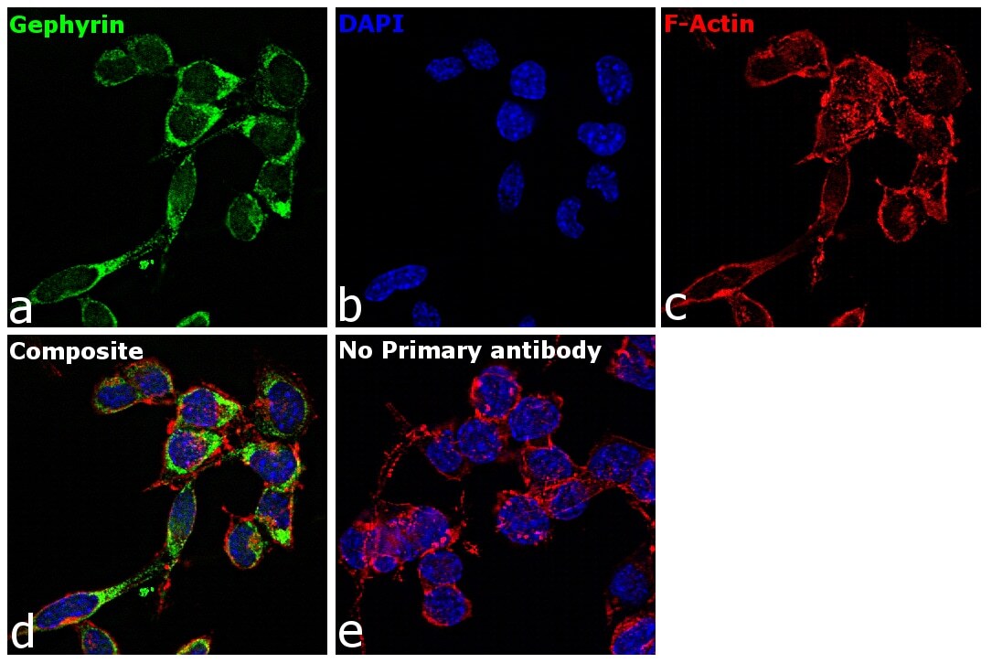
- Immunofluorescence analysis of Gephyrin was performed using 70% confluent log phase Neuro-2a cells. The cells were fixed with 4% paraformaldehyde for 10 minutes, permeabilized with 0.1% Triton™ X-100 for 15 minutes, and blocked with 1% BSA for 1 hour at room temperature. The cells were labeled with Gephyrin Rabbit Polyclonal Antibody (Product # PA5-29036) at 5 microgram/mL in 0.1% BSA, incubated at 4 degree Celsius overnight and then labeled with Goat anti-Rabbit IgG (Heavy Chain) Superclonal™ Secondary Antibody, Alexa Fluor® 488 conjugate (Product # A27034) at a dilution of 1:2000 for 45 minutes at room temperature (Panel a: green). Nuclei (Panel b: blue) were stained with ProLong™ Diamond Antifade Mountant with DAPI (Product # P36962). F-actin (Panel c: red) was stained with Rhodamine Phalloidin (Product # R415). Panel d represents the merged image showing cytoplasmic localization. Panel e represents control cells with no primary antibody to assess background. The images were captured at 60X magnification.
- Submitted by
- Invitrogen Antibodies (provider)
- Main image
- Experimental details
- Immunocytochemistry-Immunofluorescence analysis of Gephyrin was performed in DIV10 rat E18 primary hippocampal neurons fixed in 4% paraformaldehyde at RT for 15 min. Green: Gephyrin Polyclonal Antibody (Product # PA5-29036) diluted at 1:500. Red: beta Tubulin 3/ Tuj1, stained by beta Tubulin 3/ Tuj1 antibody. Blue: Fluoroshield with DAPI.
Supportive validation
- Submitted by
- Invitrogen Antibodies (provider)
- Main image
- Experimental details
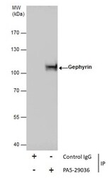
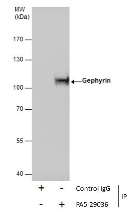
- Immunoprecipitation of Gephyrin was performed in A431 whole cell extracts using 5 µg of Gephyrin Polyclonal Antibody (Product # PA5-29036). Samples were transferred to a membrane and probed with Gephyrin Polyclonal Antibody as a primary antibody and an HRP-conjugated anti-Rabbit IgG was used as a secondary antibody.
Supportive validation
- Submitted by
- Invitrogen Antibodies (provider)
- Main image
- Experimental details
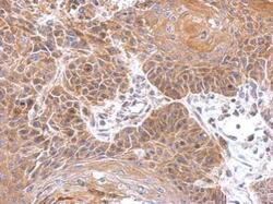
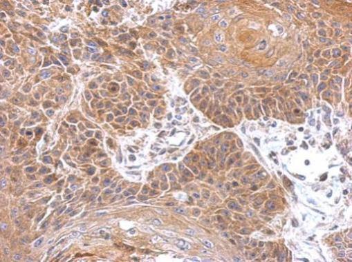
- Immunohistochemical analysis of paraffin-embedded mouse brain, using Gephyrin (Product # PA5-29036) antibody at 1:500 dilution. Antigen Retrieval: EDTA based buffer, pH 8.0, 15 min.
- Submitted by
- Invitrogen Antibodies (provider)
- Main image
- Experimental details
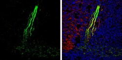
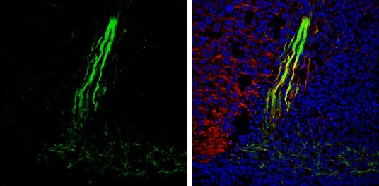
- Immunohistochemistry (Frozen) analysis of Gephyrin was performed in frozen sectioned E13.5 Rat brain tissue using Gephyrin Polyclonal Antibody (Product # PA5-29036) at a dilution of 1:250 (Green). Red: beta Tubulin 3/ TUJ1, a mature neuron marker, stained by beta Tubulin 3/ TUJ1 antibody diluted at 1:500. Blue: Fluoroshield with DAPI.
Supportive validation
- Submitted by
- Invitrogen Antibodies (provider)
- Main image

- Experimental details
- Immunoprecipitation of Gephyrin was performed in A431 whole cell extracts using 5 µg of Gephyrin Polyclonal Antibody (Product # PA5-29036). Samples were transferred to a membrane and probed with Gephyrin Polyclonal Antibody as a primary antibody and an HRP-conjugated anti-Rabbit IgG was used as a secondary antibody.
