 Explore
 Validate
 Learn
Learn Western blot
 ELISA
ELISAAntibody data
- Antibody Data
- Antigen structure
- References [1]
- Comments [0]
- Validations
- ELISA [1]
- Immunocytochemistry [2]
- Chromatin Immunoprecipitation [2]
- Other assay [1]
Submit
Validation data
Reference
Comment
Report error
- Product number
- MA3-054 - Provider product page

- Provider
- Invitrogen Antibodies
- Product name
- HP1 gamma Monoclonal Antibody (2MOD-1G6)
- Antibody type
- Monoclonal
- Antigen
- Recombinant full-length protein
- Description
- MA3-054 detects HP1-gamma in human, mouse and rat samples. MA3-054 has been succesfully used in western blot, ELISA, immunofluorescence and ChIP applications. Immunogen is a HP1-gamma recombinant mouse
- Reactivity
- Human, Mouse, Rat
- Host
- Mouse
- Isotype
- IgG
- Antibody clone number
- 2MOD-1G6
- Vial size
- 50 μL
- Concentration
- Conc. Not Determined
- Storage
- -20°C, Avoid Freeze/Thaw Cycles
Submitted references TET1 regulates gene expression and repression of endogenous retroviruses independent of DNA demethylation.
Stolz P, Mantero AS, Tvardovskiy A, Ugur E, Wange LE, Mulholland CB, Cheng Y, Wierer M, Enard W, Schneider R, Bartke T, Leonhardt H, Elsässer SJ, Bultmann S
Nucleic acids research 2022 Aug 26;50(15):8491-8511
Nucleic acids research 2022 Aug 26;50(15):8491-8511
No comments: Submit comment
Supportive validation
- Submitted by
- Invitrogen Antibodies (provider)
- Main image
- Experimental details
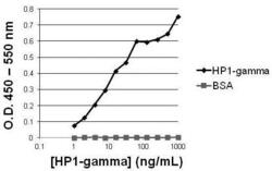
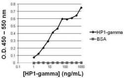
- ELISA analysis of HP1-gamma was performed by coating wells of a 96-well plate with 100 µL per well of recombinant HP1-gamma protein or BSA (Product # 23209) diluted in carbonate/bicarbonate buffer (Product # 28382), at a concentration of 1 µg/mL. Wells of the plate were washed, blocked with Superblock blocking buffer (Product # 37536), and incubated with 100 µL per well of a mouse anti-HP1-gamma monoclonal antibody (Product # MA3-054) starting at a concentration of 1 µg/mL and serially diluting 2-fold to a concentration of 1 ng/mL for 1 hour at 37°C. The plate was washed, then incubated with 100 µL per well of goat anti-mouse IgG SuperclonalTM secondary antibody (Product # A28174) at a dilution of 1:10,000 for 1 hour at 37°C followed by washing. Detection was performed using 1-Step Ultra TMB substrate (Product # 34028) for 5 minutes at room temperature in the dark. The reaction was stopped with 0.16M sulfuric acid, and absorbances were read on a spectrophotometer at 450-550 nm.

Supportive validation
- Submitted by
- Invitrogen Antibodies (provider)
- Main image
- Experimental details
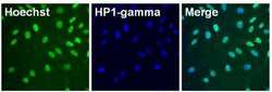
- Immunofluorescent analysis of HP1-gamma (green) in HeLa cells. The cells were fixed with formalin for 15 minutes, permeabilized with 1X Permeablilization buffer (Product # 8408400) for 15 minutes, and blocked with 1% Blocker BSA (Product # 37525) for 15 minutes at room temperature. Cells were stained with HP1-gamma monoclonal antibody (Product # MA3-054) at a dilution of 1:250 in blocking buffer for one hour at room temperature, washed with 1X TBS Tween 20 Buffer (Product # 28360), followed by incubation with DyLight 488 goat anti-mouse IgG secondary antibody (green, Product # A28175) at a dilution of 1:2000 and Hoechst 33342 dye (blue, Product # 62249) at a dilution of 1:5000 for 30 minutes at room temperature. Images were taken on a Thermo Scientific ToxInsight at 20X magnification.

- Submitted by
- Invitrogen Antibodies (provider)
- Main image

- Experimental details
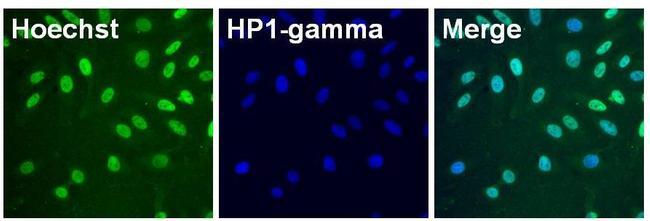
- Immunofluorescent analysis of HP1-gamma (green) in HeLa cells. The cells were fixed with formalin for 15 minutes, permeabilized with 1X Permeablilization buffer (Product # 8408400) for 15 minutes, and blocked with 1% Blocker BSA (Product # 37525) for 15 minutes at room temperature. Cells were stained with HP1-gamma monoclonal antibody (Product # MA3-054) at a dilution of 1:250 in blocking buffer for one hour at room temperature, washed with 1X TBS Tween 20 Buffer (Product # 28360), followed by incubation with DyLight 488 goat anti-mouse IgG secondary antibody (green, Product # A28175) at a dilution of 1:2000 and Hoechst 33342 dye (blue, Product # 62249) at a dilution of 1:5000 for 30 minutes at room temperature. Images were taken on a Thermo Scientific ToxInsight at 20X magnification.
Supportive validation
- Submitted by
- Invitrogen Antibodies (provider)
- Main image
- Experimental details
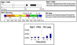
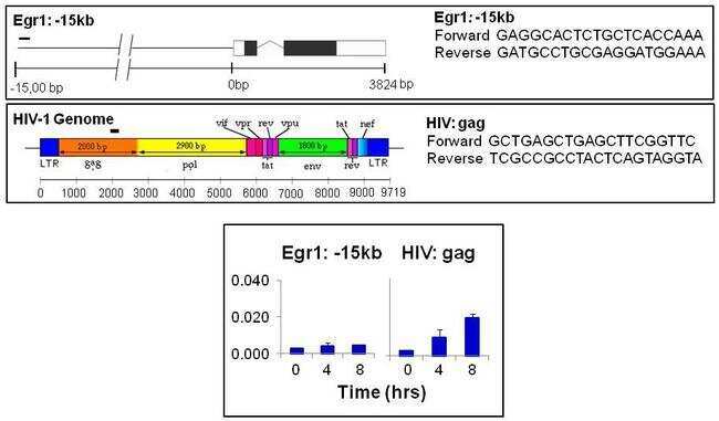
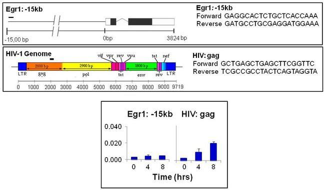
- Chromatin immunoprecipitation analysis of HIV1 HP1-gamma was performed using cross-linked chromatin from Human 5A8 J-lat T lymphocytes culture latently infected with HIV1 and treated with 10 µg/mL PHA (phytohemaglutin) for 0, 4, and 8 hours. Immunoprecipitation was performed using a multiplex microplate Matrix ChIP assay (see reference for Matrix ChIP protocol: http://www.ncbi.nlm.nih.gov/pubmed/22098709) with 1.0 µL/100 µL well volume of HP1-gamma monoclonal antibody (Product # MA3-054). Chromatin aliquots from ~1 x 10⁵ cells were used per ChIP pull-down. Quantitative PCR data were done in quadruplicate using 1 µL of eluted DNA in 2 µL SYBR real-time PCR reactions containing primers shown to amplify exon-1 of the Egr1 gene or HIV gag gene. PCR calibration curves were generated for each primer pair from a dilution series of sheared total genomic DNA. Quantitation of immunoprecipitated chromatin is presented as signal relative to the total amount of input chromatin. Results represent the mean +/- SEM for three experiments. Schematic representations of the Egr1 and HIV1 gag locus are shown above the data where boxes represent exons (black boxes = translated regions, white boxes = untranslated regions), the zigzag line represents an intron, and the straight line represents upstream sequence. Regions amplified by the primers are represented by black bars. Data courtesy of the Innovators Program.
- Submitted by
- Invitrogen Antibodies (provider)
- Main image
- Experimental details
- Chromatin immunoprecipitation analysis of HIV1 HP1-gamma was performed using cross-linked chromatin from Human 5A8 J-lat T lymphocytes culture latently infected with HIV1 and treated with 10 µg/mL PHA (phytohemaglutin) for 0, 4, and 8 hours. Immunoprecipitation was performed using a multiplex microplate Matrix ChIP assay (see reference for Matrix ChIP protocol: http://www.ncbi.nlm.nih.gov/pubmed/22098709) with 1.0 µL/100 µL well volume of HP1-gamma monoclonal antibody (Product # MA3-054). Chromatin aliquots from ~1 x 10⁵ cells were used per ChIP pull-down. Quantitative PCR data were done in quadruplicate using 1 µL of eluted DNA in 2 µL SYBR real-time PCR reactions containing primers shown to amplify exon-1 of the Egr1 gene or HIV gag gene. PCR calibration curves were generated for each primer pair from a dilution series of sheared total genomic DNA. Quantitation of immunoprecipitated chromatin is presented as signal relative to the total amount of input chromatin. Results represent the mean +/- SEM for three experiments. Schematic representations of the Egr1 and HIV1 gag locus are shown above the data where boxes represent exons (black boxes = translated regions, white boxes = untranslated regions), the zigzag line represents an intron, and the straight line represents upstream sequence. Regions amplified by the primers are represented by black bars. Data courtesy of the Innovators Program.
Supportive validation
- Submitted by
- Invitrogen Antibodies (provider)
- Main image
- Experimental details
- ELISA analysis of HP1-gamma was performed by coating wells of a 96-well plate with 100 µL per well of recombinant HP1-gamma protein or BSA (Product # 23209) diluted in carbonate/bicarbonate buffer (Product # 28382), at a concentration of 1 µg/mL. Wells of the plate were washed, blocked with Superblock blocking buffer (Product # 37536), and incubated with 100 µL per well of a mouse anti-HP1-gamma monoclonal antibody (Product # MA3-054) starting at a concentration of 1 µg/mL and serially diluting 2-fold to a concentration of 1 ng/mL for 1 hour at 37°C. The plate was washed, then incubated with 100 µL per well of goat anti-mouse IgG SuperclonalTM secondary antibody (Product # A28174) at a dilution of 1:10,000 for 1 hour at 37°C followed by washing. Detection was performed using 1-Step Ultra TMB substrate (Product # 34028) for 5 minutes at room temperature in the dark. The reaction was stopped with 0.16M sulfuric acid, and absorbances were read on a spectrophotometer at 450-550 nm.
