 Explore
 Validate
 Learn
Learn Western blot
Antibody data
- Antibody Data
- Antigen structure
- References [2]
- Comments [0]
- Validations
- Western blot [2]
- Immunocytochemistry [2]
Submit
Validation data
Reference
Comment
Report error
- Product number
- AF3619 - Provider product page

- Provider
- R&D Systems
- Product name
- Human FoxJ1 Antibody
- Antibody type
- Polyclonal
- Description
- Antigen Affinity-purified. Detects human FoxJ1 in direct ELISAs and Western blots.
- Reactivity
- Human
- Host
- Goat
- Conjugate
- Unconjugated
- Antigen sequence
Q92949- Isotype
- IgG
- Vial size
- 100 ug
- Concentration
- LYOPH
- Storage
- Use a manual defrost freezer and avoid repeated freeze-thaw cycles. 12 months from date of receipt, -20 to -70 °C as supplied. 1 month, 2 to 8 °C under sterile conditions after reconstitution. 6 months, -20 to -70 °C under sterile conditions after reconstitution.
Submitted references Gefitinib, an EGFR Tyrosine Kinase inhibitor, Prevents Smoke-Mediated Ciliated Airway Epithelial Cell Loss and Promotes Their Recovery.
Hyaluronan fragments/CD44 mediate oxidative stress-induced MUC5B up-regulation in airway epithelium.
Valencia-Gattas M, Conner GE, Fregien NL
PloS one 2016;11(8):e0160216
PloS one 2016;11(8):e0160216
Hyaluronan fragments/CD44 mediate oxidative stress-induced MUC5B up-regulation in airway epithelium.
Casalino-Matsuda SM, Monzon ME, Day AJ, Forteza RM
American journal of respiratory cell and molecular biology 2009 Mar;40(3):277-85
American journal of respiratory cell and molecular biology 2009 Mar;40(3):277-85
No comments: Submit comment
Supportive validation
- Submitted by
- R&D Systems (provider)
- Main image
- Experimental details
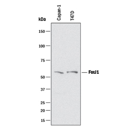
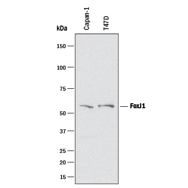
- Detection of Human FoxJ1 by Western Blot. Western blot shows lysates of Capan-1 human pancreatic adenocarcinoma cell line and T47D human breast cancer cell line. PVDF membrane was probed with 1 µg/mL of Goat Anti-Human FoxJ1 Antigen Affinity-purified Polyclonal Antibody (Catalog # AF3619) followed by HRP-conjugated Anti-Goat IgG Secondary Antibody (Catalog # HAF017). A specific band was detected for FoxJ1 at approximately 55 kDa (as indicated). This experiment was conducted under reducing conditions and using Immunoblot Buffer Group 1.
- Submitted by
- R&D Systems (provider)
- Main image
- Experimental details
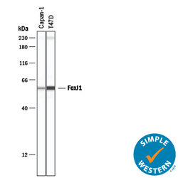
- Detection of Human FoxJ1 by Simple WesternTM. Simple Western lane view shows lysates of Capan-1 human pancreatic adenocarcinoma cell line and T47D human breast cancer cell line, loaded at 0.2 mg/mL. A specific band was detected for FoxJ1 at approximately 58 kDa (as indicated) using 10 µg/mL of Goat Anti-Human FoxJ1 Antigen Affinity-purified Polyclonal Antibody (Catalog # AF3619) followed by 1:50 dilution of HRP-conjugated Anti-Goat IgG Secondary Antibody (Catalog # HAF019). This experiment was conducted under reducing conditions and using the 12-230 kDa separation system. Non-specific interaction with the 230 kDa Simple Western standard may be seen with this antibody.

Supportive validation
- Submitted by
- R&D Systems (provider)
- Main image
- Experimental details
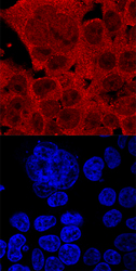
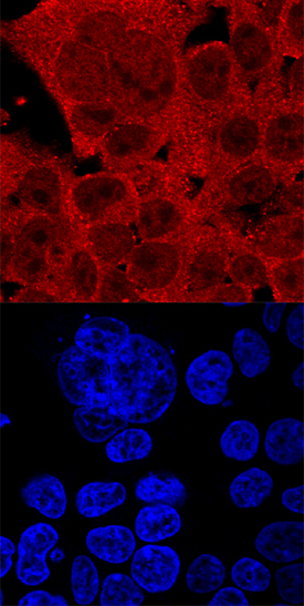
- FoxJ1 in HEK293 Human Cell Line. FoxJ1 was detected in immersion fixed HEK293 human embryonic kidney cell line using Goat Anti-Human FoxJ1 Antigen Affinity-purified Polyclonal Antibody (Catalog # AF3619) at 10 µg/mL for 3 hours at room temperature. Cells were stained using the NorthernLights™ 557-conjugated Anti-Goat IgG Secondary Antibody (red, upper panel; Catalog # NL001) and counterstained with DAPI (blue, lower panel). Specific staining was localized to nuclei and cytoplasm. View our protocol for Fluorescent ICC Staining of Cells on Coverslips.
- Submitted by
- R&D Systems (provider)
- Main image
- Experimental details
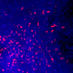
- FoxJ1 in Early Proximal Lung Progenitors. FoxJ1 was detected in immersion fixed BGO1V human embryonic stem cells differentiated to early proximal lung progenitors using Goat Anti-Human FoxJ1 Antigen Affinity-purified Polyclonal Antibody (Catalog # AF3619) at 10 µg/mL for 3 hours at room temperature. Cells were stained using the NorthernLights™ 557-conjugated Anti-Goat IgG Secondary Antibody (red; Catalog # NL001) and counterstained with DAPI (blue). Specific staining was localized to nuclei. View our protocol for Fluorescent ICC Staining of Stem Cells on Coverslips.
