Explore
 Validate
 Learn
Learn Western blot
 Immunocytochemistry
Antibody data
- Antibody Data
- Antigen structure
- References [0]
- Comments [0]
- Validations
- Immunocytochemistry [2]
Submit
Validation data
Reference
Comment
Report error
- Product number
- 720280 - Provider product page

- Provider
- Invitrogen Antibodies
- Product name
- mGluR4 Polyclonal Antibody
- Antibody type
- Polyclonal
- Antigen
- Synthetic peptide
- Reactivity
- Human
- Host
- Rabbit
- Isotype
- IgG
- Vial size
- 100 µg
- Concentration
- 0.5 mg/mL
- Storage
- Store at 4°C short term. For long term storage, store at -20°C, avoiding freeze/thaw cycles.
No comments: Submit comment
Supportive validation
- Submitted by
- Invitrogen Antibodies (provider)
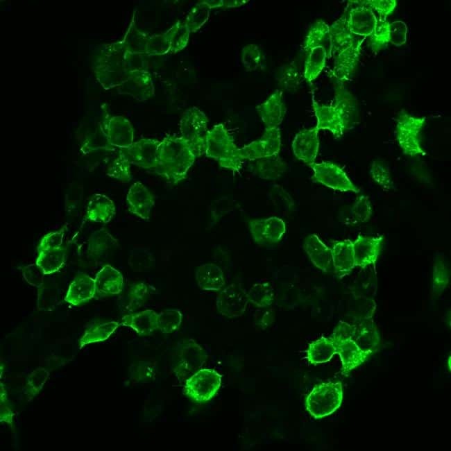
- Main image
- Experimental details
- HEK 293 cells were grown on poly-L-lysine-coated coverslips overnight. Cells were then transiently transfected with mGLUR4. After 24 h, cells were fixed with 4% paraformaldehyde and 0.2% picric acid in phosphate buffer (pH 6.9) for 30 min at room temperature and washed several times with phosphate buffer. Specimens were permeabilized with ice-cold methanol and then incubated with Anti-mGLUR4 Rabbit Polyclonal Antibody (Product # 720280 at a concentration of 2 µg/mL). Cells were then incubated with Alexa488-conjugated secondary antibody for 2 h at room temperature, mounted and examined using a laser scanning confocal microscope. Note, strong staining of transfected cells and no staining of adjacent non-transfected cells.

- Submitted by
- Invitrogen Antibodies (provider)
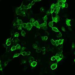
- Main image

- Experimental details
- HEK 293 cells were grown on poly-L-lysine-coated coverslips overnight. Cells were then transiently transfected with mGLUR4. After 24 h, cells were fixed with 4% paraformaldehyde and 0.2% picric acid in phosphate buffer (pH 6.9) for 30 min at room temperature and washed several times with phosphate buffer. Specimens were permeabilized with ice-cold methanol and then incubated with Anti-mGLUR4 Rabbit Polyclonal Antibody (Product # 720280 at a concentration of 2 µg/mL). Cells were then incubated with Alexa488-conjugated secondary antibody for 2 h at room temperature, mounted and examined using a laser scanning confocal microscope. Note, strong staining of transfected cells and no staining of adjacent non-transfected cells.