Explore
 Validate
 Learn
Learn Western blot
Antibody data
- Antibody Data
- Antigen structure
- References [3]
- Comments [0]
- Validations
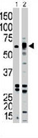
- Western blot [1]
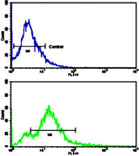
- Flow cytometry [1]
Submit
Validation data
Reference
Comment
Report error
- Product number
- PAB3467 - Provider product page

- Provider
- Abnova Corporation
- Proper citation
- Abnova Corporation Cat#PAB3467, RRID:AB_1571122
- Product name
- ACVR1B polyclonal antibody
- Antibody type
- Polyclonal
- Description
- Rabbit polyclonal antibody raised against synthetic peptide of ACVR1B.
- Storage
- Store at 4°C. For long term storage store at -20°C.Aliquot to avoid repeated freezing and thawing.
Submitted references Identification of a functional binding site for activin on the type I receptor ALK4.
Activin signaling through type IB activin receptor stimulates aromatase activity in the ovarian granulosa cell-like human granulosa (KGN) cells.
Overexpression of wild-type activin receptor alk4-1 restores activin antiproliferative effects in human pituitary tumor cells.
Harrison CA, Gray PC, Koerber SC, Fischer W, Vale W
The Journal of biological chemistry 2003 Jun 6;278(23):21129-35
The Journal of biological chemistry 2003 Jun 6;278(23):21129-35
Activin signaling through type IB activin receptor stimulates aromatase activity in the ovarian granulosa cell-like human granulosa (KGN) cells.
Mukasa C, Nomura M, Tanaka T, Tanaka K, Nishi Y, Okabe T, Goto K, Yanase T, Nawata H
Endocrinology 2003 Apr;144(4):1603-11
Endocrinology 2003 Apr;144(4):1603-11
Overexpression of wild-type activin receptor alk4-1 restores activin antiproliferative effects in human pituitary tumor cells.
Danila DC, Zhang X, Zhou Y, Haidar JN, Klibanski A
The Journal of clinical endocrinology and metabolism 2002 Oct;87(10):4741-6
The Journal of clinical endocrinology and metabolism 2002 Oct;87(10):4741-6
No comments: Submit comment
Supportive validation
- Submitted by
- Abnova Corporation (provider)
- Main image

- Experimental details
- The ACVR1B polyclonal antibody (Cat # PAB3467) is used in Western blot to detect ACVR1B in Jurkat (1) and mouse kidney (2) tissue lysates.
Supportive validation
- Submitted by
- Abnova Corporation (provider)
- Main image

- Experimental details
- Flow cytometric analysis of 293 cells using ACVR1B polyclonal antibody (Cat # PAB3467)(bottom histogram) compared to a negative control cell (top histogram).FITC-conjugated goat-anti-rabbit secondary antibodies were used for the analysis.
- Validation comment
- Flow Cytometry