 Explore
 Validate
 Learn
Learn Western blot
 ELISA
ELISAAntibody data
- Antibody Data
- Antigen structure
- References [0]
- Comments [0]
- Validations
- Western blot [3]
Submit
Validation data
Reference
Comment
Report error
- Product number
- MA5-17209 - Provider product page

- Provider
- Invitrogen Antibodies
- Product name
- Anti-CDK7 Monoclonal Antibody (3E3)
- Antibody type
- Monoclonal
- Antigen
- Recombinant full-length protein
- Description
- A suggested positive control for this product is HeLa cells.
- Reactivity
- Human
- Host
- Mouse
- Isotype
- IgG
- Antibody clone number
- 3E3
- Vial size
- 100 µL
- Concentration
- 1 mg/mL
- Storage
- -20°C
No comments: Submit comment
Supportive validation
- Submitted by
- Invitrogen Antibodies (provider)
- Main image
- Experimental details
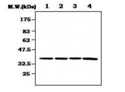
- Western blot analysis of CDK7 using anti-CDK7 monoclonal antibody (Product # MA5-17209) on (Lane 1) HeLa Cell lysate, (Lane 2) Jurket Cell lysate, (Lane 3) SK-N-MC Cell lysate and K562 Cell lysate.

- Submitted by
- Invitrogen Antibodies (provider)
- Main image
- Experimental details
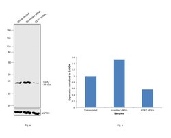
- Knockdown of CDK7 was achieved by transfecting A-431 with CDK7 specific siRNAs (Silencer® select Product # s2830). Western blot analysis (Fig. a) was performed using Whole cell extracts from the CDK7 knockdown cells (lane 3), non-specific scrambled siRNA transfected cells (lane 2) and untransfected cells (lane 1). The blot was probed with CDK7 Monoclonal Antibody (3E3) (Product # MA5-17209, 0.4 ug/ml) and Goat anti-Mouse IgG (H+L) Superclonal™ Recombinant Secondary Antibody, HRP (Product # A28177, 1:4000 dilution). Densitometric analysis of this western blot is shown in histogram (Fig. b). Decrease in signal upon siRNA mediated knock down confirms that antibody is specific to CDK7.

- Submitted by
- Invitrogen Antibodies (provider)
- Main image
- Experimental details
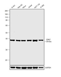
- Western blot was performed using Anti-CDK7 Monoclonal Antibody (3E3) (Product # MA5-17209) and a 39 kDa band corresponding to CDK7 was observed across cell lines tested. Whole cell extracts (30 µg lysate) of A-431 (Lane 1), Hep G2 (Lane 2), HeLa (Lane 3), Jurkat (Lane 4), HCT 116 (Lane 5) and K-562 (Lane 6) were electrophoresed using NuPAGE™ 4-12% Bis-Tris Protein Gel (Product # NP0322BOX). Resolved proteins were then transferred onto a nitrocellulose membrane (Product # IB23001) by iBlot® 2 Dry Blotting System (Product # IB21001). The blot was probed with the primary antibody (0.4 ug/ml) and detected by chemiluminescence with Goat anti-Mouse IgG (H+L) Superclonal™ Recombinant Secondary Antibody, HRP (Product # A28177, 1:4000 dilution), 1:4000 dilution) using the iBright FL 1000 (Product # A32752). Chemiluminescent detection was performed using Novex® ECL Chemiluminescent Substrate Reagent Kit (Product # WP20005).
