Explore
 Validate
 Learn
Learn Western blot
 Immunoprecipitation
Antibody data
- Antibody Data
- Antigen structure
- References [1]
- Comments [0]
- Validations
- Immunoprecipitation [1]
- Immunohistochemistry [1]
- Other assay [1]
Submit
Validation data
Reference
Comment
Report error
- Product number
- PA5-34791 - Provider product page

- Provider
- Invitrogen Antibodies
- Product name
- CDK7 Polyclonal Antibody
- Antibody type
- Polyclonal
- Antigen
- Recombinant full-length protein
- Description
- Recommended positive controls: 293T, A431, A375, Neuro2A, GL261, C8D30, NIH-3T3, BCL-1, Raw264.7, C2C12. Predicted reactivity: Mouse (95%), Zebrafish (85%), Xenopus laevis (86%), Pig (97%), Rhesus Monkey (99%), Bovine (96%). IHC notes, Requires antigen retrieval using heat mediated 10mM Citrate buffer (pH6.0) or Tris-EDTA buffer (pH8.0) Store product as a concentrated solution. Centrifuge briefly prior to opening the vial.
- Reactivity
- Human, Mouse
- Host
- Rabbit
- Isotype
- IgG
- Vial size
- 100 μL
- Concentration
- 0.96 mg/mL
- Storage
- Store at 4°C short term. For long term storage, store at -20°C, avoiding freeze/thaw cycles.
Submitted references Cell-Cycle Proteins Control Production of Neutrophil Extracellular Traps.
Amulic B, Knackstedt SL, Abu Abed U, Deigendesch N, Harbort CJ, Caffrey BE, Brinkmann V, Heppner FL, Hinds PW, Zychlinsky A
Developmental cell 2017 Nov 20;43(4):449-462.e5
Developmental cell 2017 Nov 20;43(4):449-462.e5
No comments: Submit comment
Supportive validation
- Submitted by
- Invitrogen Antibodies (provider)
- Main image

- Experimental details
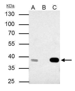
- CDK7 Polyclonal Antibody immunoprecipitates CDK7 protein in IP experiments. IP samples: 293T whole cell extract. A. 40 µg 293T whole cell extract. B. Control with 4 µg of preimmune Rabbit IgG. C. Immunoprecipitation of CDK7 protein by 4 µg CDK7 Polyclonal Antibody (Product # PA5-34791). 10 % SDS-PAGE. The immunoprecipitated CDK7 protein was detected by CDK7 Polyclonal Antibody (Product # PA5-34791) diluted at 1:500.

Supportive validation
- Submitted by
- Invitrogen Antibodies (provider)
- Main image
- Experimental details
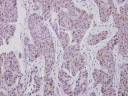
- Immunohistochemical analysis of paraffin-embedded human breast cancer, using CDK7 (Product # PA5-34791) antibody at 1:250 dilution. Antigen Retrieval: EDTA based buffer, pH 8.0, 15 min.

Supportive validation
- Submitted by
- Invitrogen Antibodies (provider)
- Main image
- Experimental details
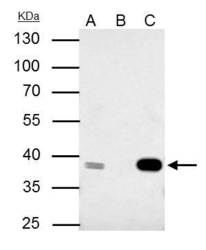
- CDK7 Polyclonal Antibody immunoprecipitates CDK7 protein in IP experiments. IP samples: 293T whole cell extract. A. 40 µg 293T whole cell extract. B. Control with 4 µg of preimmune Rabbit IgG. C. Immunoprecipitation of CDK7 protein by 4 µg CDK7 Polyclonal Antibody (Product # PA5-34791). 10 % SDS-PAGE. The immunoprecipitated CDK7 protein was detected by CDK7 Polyclonal Antibody (Product # PA5-34791) diluted at 1:500.