 Explore
 Validate
 Learn
Learn Western blot
 Immunoprecipitation
Antibody data
- Antibody Data
- Antigen structure
- References [0]
- Comments [0]
- Validations
- Immunoprecipitation [1]
- Immunohistochemistry [1]
- Other assay [2]
Submit
Validation data
Reference
Comment
Report error
- Product number
- PA5-31554 - Provider product page

- Provider
- Invitrogen Antibodies
- Product name
- RAD50 Polyclonal Antibody
- Antibody type
- Polyclonal
- Antigen
- Recombinant full-length protein
- Description
- Recommended positive controls: HeLa, NT2D1, Neuro2A, GL261, PC-12. Predicted reactivity: Mouse (92%), Rat (91%), Rabbit (92%), Bovine (93%). Store product as a concentrated solution. Centrifuge briefly prior to opening the vial.
- Reactivity
- Human, Mouse, Rat
- Host
- Rabbit
- Isotype
- IgG
- Vial size
- 100 μL
- Concentration
- 0.78 mg/mL
- Storage
- Store at 4°C short term. For long term storage, store at -20°C, avoiding freeze/thaw cycles.
No comments: Submit comment
Supportive validation
- Submitted by
- Invitrogen Antibodies (provider)
- Main image

- Experimental details
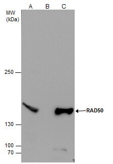
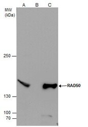
- RAD50 Polyclonal Antibody immunoprecipitates Rad50 protein in IP experiments. IP samples: NT2D1 whole cell extract. A. 50 µg NT2D1 whole cell extract. B. Control with 4 µg of preimmune Rabbit IgG. C. Immunoprecipitation of Rad50 protein by 4 µg RAD50 Polyclonal Antibody (Product # PA5-31554). 5 % SDS-PAGE. The immunoprecipitated Rad50 protein was detected by RAD50 Polyclonal Antibody (Product # PA5-31554) diluted at 1:500.
Supportive validation
- Submitted by
- Invitrogen Antibodies (provider)
- Main image

- Experimental details
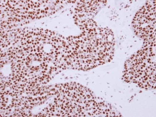
- Immunohistochemical analysis of paraffin-embedded human lung carcinoma, using Rad50 (Product # PA5-31554) antibody at 1:250 dilution. Antigen Retrieval: EDTA based buffer, pH 8.0, 15 min.
Supportive validation
- Submitted by
- Invitrogen Antibodies (provider)
- Main image
- Experimental details
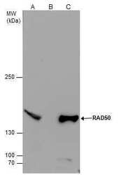
- RAD50 Polyclonal Antibody immunoprecipitates Rad50 protein in IP experiments. IP samples: NT2D1 whole cell extract. A. 50 µg NT2D1 whole cell extract. B. Control with 4 µg of preimmune Rabbit IgG. C. Immunoprecipitation of Rad50 protein by 4 µg RAD50 Polyclonal Antibody (Product # PA5-31554). 5 % SDS-PAGE. The immunoprecipitated Rad50 protein was detected by RAD50 Polyclonal Antibody (Product # PA5-31554) diluted at 1:500.
- Submitted by
- Invitrogen Antibodies (provider)
- Main image
- Experimental details
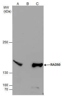
- RAD50 Polyclonal Antibody immunoprecipitates Rad50 protein in IP experiments. IP samples: NT2D1 whole cell extract. A. 50 µg NT2D1 whole cell extract. B. Control with 4 µg of preimmune Rabbit IgG. C. Immunoprecipitation of Rad50 protein by 4 µg RAD50 Polyclonal Antibody (Product # PA5-31554). 5 % SDS-PAGE. The immunoprecipitated Rad50 protein was detected by RAD50 Polyclonal Antibody (Product # PA5-31554) diluted at 1:500.
