Explore
 Validate
 Learn
LearnSTJ11101280
antibody from St John's Laboratory
Targeting: CDK9
C-2k, CDC2L4, PITALRE, TAK
 Western blot
 Immunocytochemistry Immunoprecipitation Immunohistochemistry Chromatin Immunoprecipitation
Antibody data
- Antibody Data
- Antigen structure
- References [0]
- Comments [0]
- Validations
- Western blot [1]
- Immunohistochemistry [5]
- Chromatin Immunoprecipitation [2]
Submit
Validation data
Reference
Comment
Report error
- Product number
- STJ11101280 - Provider product page

- Provider
- St John's Laboratory
- Product name
- Anti-CDK9 antibody [ARC0527] (STJ11101280)
- Antibody type
- Monoclonal
- Description
- Rabbit monoclonal antibody anti-CDK9 is suitable for use in Western Blot, Immunohistochemistry, Immunofluorescence and Immunoprecipitation.
- Reactivity
- Human, Mouse, Rat
- Host
- Rabbit
- Conjugate
- Unconjugated
- Antigen sequence
NA- Epitope
- NA
- Isotype
- IgG
- Antibody clone number
- NA
- Vial size
- NA
- Concentration
- NA
- Storage
- Store in a freezer at-20°C and avoid freeze-thaw cycles.
- Handling
- NA
No comments: Submit comment
Supportive validation
- Submitted by
- St John's Laboratory (provider)
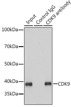
- Main image
- Experimental details
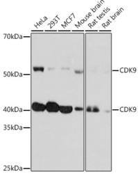
- Western blot analysis of extracts of various cell lines, using CDK9 rabbit monoclonal antibody (STJ11101280) at 1:1000 dilution. Secondary antibody: HRP Goat Anti-rabbit IgG (H+L) at 1:10000 dilution. Lysates/proteins: 25ug per lane. Blocking buffer: 3% nonfat dry milk in TBST. Detection: ECL Basic Kit. Exposure time: 3min.
- Sample type
- NA
- Validation comment
- NA
- Primary Ab dilution
- NA
- Other comments
- NA
- Secondary Ab
- NA
- Secondary Ab dilution
- NA
- Protocol
- NA
Supportive validation
Supportive validation
Supportive validation
Supportive validation
Supportive validation
- Submitted by
- St John's Laboratory (provider)
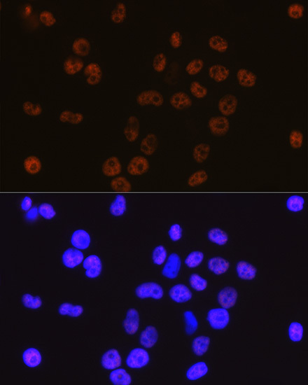
- Main image
- Experimental details
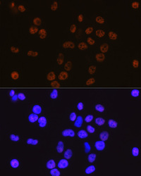
- Immunofluorescence analysis of HeLa cells using CDK9 rabbit monoclonal antibody (STJ11101280) at dilution of 1:100 (40x lens). Blue: DAPI for nuclear staining.
- Sample type
- NA
- Validation comment
- NA
- Primary Ab dilution
- NA
- Other comments
- NA
- Secondary Ab
- NA
- Secondary Ab dilution
- NA
- Protocol
- NA
Supportive validation
- Submitted by
- St John's Laboratory (provider)
- Main image
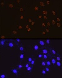
- Experimental details
- Immunofluorescence analysis of NIH-3T3 cells using CDK9 rabbit monoclonal antibody (STJ11101280) at dilution of 1:100 (40x lens). Blue: DAPI for nuclear staining.
- Sample type
- NA
- Validation comment
- NA
- Primary Ab dilution
- NA
- Other comments
- NA
- Secondary Ab
- NA
- Secondary Ab dilution
- NA
- Protocol
- NA
Supportive validation
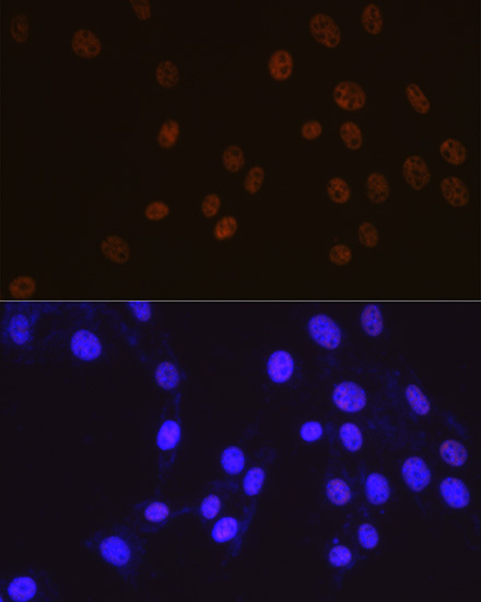
- Submitted by
- St John's Laboratory (provider)
- Main image
- Experimental details
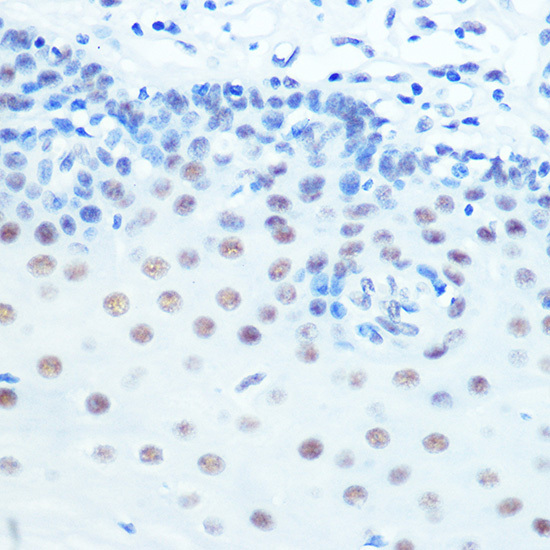
- Immunohistochemistry of paraffin-embedded rat ovary using CDK9 rabbit monoclonal antibody (STJ11101280) at dilution of 1:100 (40x lens). Perform microwave antigen retrieval with 10 mM PBS buffer pH 7. 2 before commencing with immunohistochemistry staining protocol.
- Sample type
- NA
- Validation comment
- NA
- Primary Ab dilution
- NA
- Other comments
- NA
- Secondary Ab
- NA
- Secondary Ab dilution
- NA
- Protocol
- NA
Supportive validation
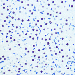
- Submitted by
- St John's Laboratory (provider)
- Main image
- Experimental details
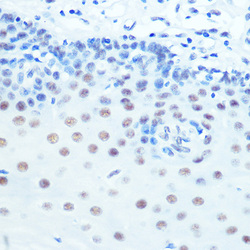
- Immunohistochemistry of paraffin-embedded human esophageal using CDK9 rabbit monoclonal antibody (STJ11101280) at dilution of 1:100 (40x lens). Perform microwave antigen retrieval with 10 mM PBS buffer pH 7. 2 before commencing with immunohistochemistry staining protocol.
- Sample type
- NA
- Validation comment
- NA
- Primary Ab dilution
- NA
- Other comments
- NA
- Secondary Ab
- NA
- Secondary Ab dilution
- NA
- Protocol
- NA
Supportive validation
- Submitted by
- St John's Laboratory (provider)
- Main image
- Experimental details
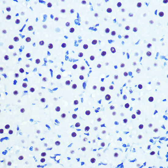
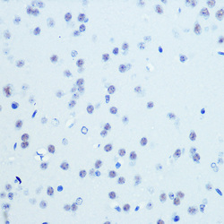
- Immunohistochemistry of paraffin-embedded mouse brain using CDK9 rabbit monoclonal antibody (STJ11101280) at dilution of 1:100 (40x lens). Perform microwave antigen retrieval with 10 mM PBS buffer pH 7. 2 before commencing with immunohistochemistry staining protocol.
- Sample type
- NA
- Validation comment
- NA
- Primary Ab dilution
- NA
- Other comments
- NA
- Secondary Ab
- NA
- Secondary Ab dilution
- NA
- Protocol
- NA

Supportive validation
Supportive validation
- Submitted by
- St John's Laboratory (provider)
- Main image

- Experimental details
- Immunoprecipitation analysis of 200ug extracts of HeLa cells using 3ug CDK9 antibody (STJ11101280). Western blot was performed from the immunoprecipitate using CDK9 antibody (STJ11101280) at a dilition of 1:1000.
- Sample type
- NA
- Validation comment
- NA
- Primary Ab dilution
- NA
- Other comments
- NA
- Secondary Ab
- NA
- Secondary Ab dilution
- NA
- Protocol
- NA
Supportive validation
- Submitted by
- St John's Laboratory (provider)
- Main image
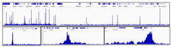
- Experimental details
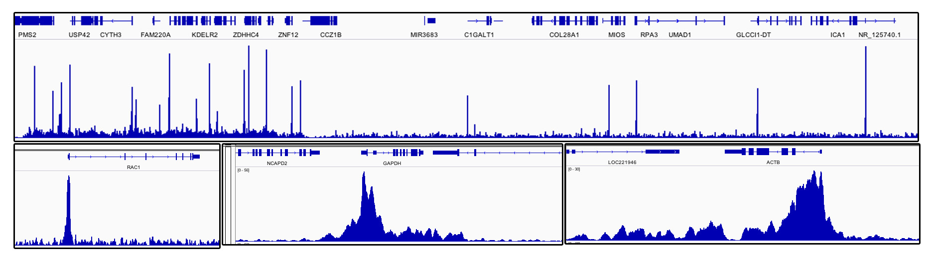
- Chromatin immunoprecipitations were performed with cross-linked chromatin from DLD-1 cells and CDK9 rabbit monoclonal antibody (STJ11101280). The ChIP sequencing results indicate the enrichment pattern of CDK9 in selected genomic region and representative gene loci (e. g. RAC1, GAPDH and ACTB) , as shown in figure.
- Sample type
- NA
- Validation comment
- NA
- Primary Ab dilution
- NA
- Other comments
- NA
- Secondary Ab
- NA
- Secondary Ab dilution
- NA
- Protocol
- NA