 Explore
 Validate
 Learn
Learn Western blot
 Immunocytochemistry
Antibody data
- Antibody Data
- Antigen structure
- References [3]
- Comments [0]
- Validations
- Immunocytochemistry [2]
- Immunoprecipitation [1]
- Immunohistochemistry [1]
- Flow cytometry [2]
- Other assay [3]
Submit
Validation data
Reference
Comment
Report error
- Product number
- MA5-14912 - Provider product page

- Provider
- Invitrogen Antibodies
- Product name
- CDK9 Monoclonal Antibody (K.513.1)
- Antibody type
- Monoclonal
- Antigen
- Synthetic peptide
- Description
- It is not recommended to aliquot this antibody.
- Reactivity
- Human, Mouse, Rat, Bovine, Canine, Hamster
- Host
- Rabbit
- Isotype
- IgG
- Antibody clone number
- K.513.1
- Vial size
- 100 μL
- Concentration
- 163 μg/mL
- Storage
- -20°C
Submitted references Epigenetic Suppression of HIV in Myeloid Cells by the BRD4-Selective Small Molecule Modulator ZL0580.
A Precision Medicine Drug Discovery Pipeline Identifies Combined CDK2 and 9 Inhibition as a Novel Therapeutic Strategy in Colorectal Cancer.
Structure-guided drug design identifies a BRD4-selective small molecule that suppresses HIV.
Alamer E, Zhong C, Liu Z, Niu Q, Long F, Guo L, Gelman BB, Soong L, Zhou J, Hu H
Journal of virology 2020 May 18;94(11)
Journal of virology 2020 May 18;94(11)
A Precision Medicine Drug Discovery Pipeline Identifies Combined CDK2 and 9 Inhibition as a Novel Therapeutic Strategy in Colorectal Cancer.
Somarelli JA, Roghani RS, Moghaddam AS, Thomas BC, Rupprecht G, Ware KE, Altunel E, Mantyh JB, Kim SY, McCall SJ, Shen X, Mantyh CR, Hsu DS
Molecular cancer therapeutics 2020 Dec;19(12):2516-2527
Molecular cancer therapeutics 2020 Dec;19(12):2516-2527
Structure-guided drug design identifies a BRD4-selective small molecule that suppresses HIV.
Niu Q, Liu Z, Alamer E, Fan X, Chen H, Endsley J, Gelman BB, Tian B, Kim JH, Michael NL, Robb ML, Ananworanich J, Zhou J, Hu H
The Journal of clinical investigation 2019 Jul 22;129(8):3361-3373
The Journal of clinical investigation 2019 Jul 22;129(8):3361-3373
No comments: Submit comment
Supportive validation
- Submitted by
- Invitrogen Antibodies (provider)
- Main image

- Experimental details
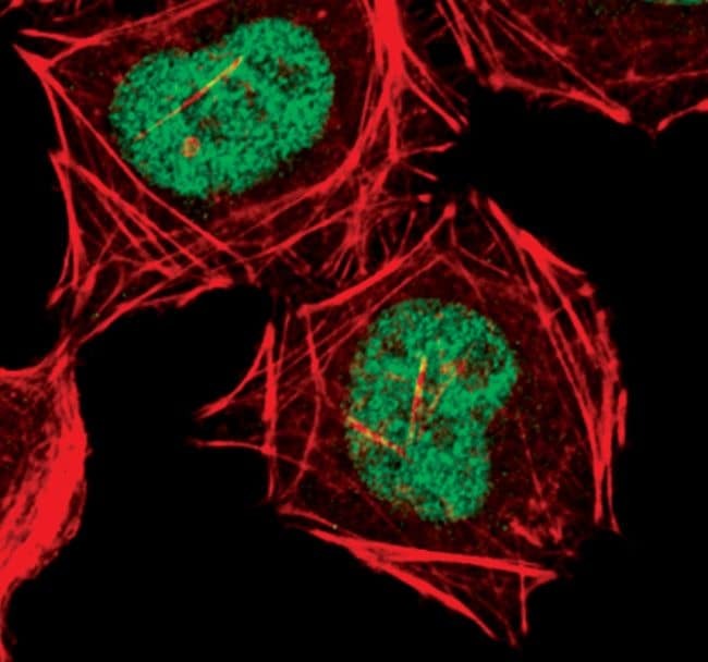
- Immunofluorescent analysis of CDK9 in HeLa cells using a CDK9 monoclonal antibody (Product # MA5-14912) (green). Actin filaments are labeled with a fluorescent red phalloidin.
- Submitted by
- Invitrogen Antibodies (provider)
- Main image
- Experimental details
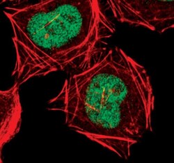
- Immunofluorescent analysis of CDK9 in HeLa cells using a CDK9 monoclonal antibody (Product # MA5-14912) (green). Actin filaments are labeled with a fluorescent red phalloidin.

Supportive validation
- Submitted by
- Invitrogen Antibodies (provider)
- Main image

- Experimental details
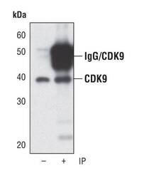
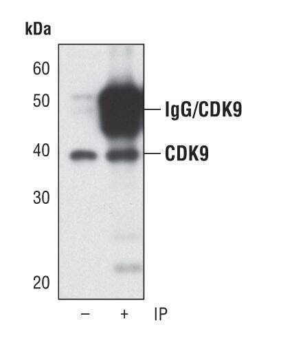
- Immunoprecipitation of CDK9 from HeLa cells using a CDK9 monoclonal antibody (Product # MA5-14912) followed by Western blot analysis using the same antibody. (Lane 1) 5% input.

Supportive validation
- Submitted by
- Invitrogen Antibodies (provider)
- Main image

- Experimental details
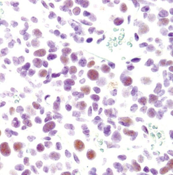
- Immunohistochemical analysis of CDK9 in paraffin-embedded K7M2 mouse syngeneic tumor using a CDK9 monoclonal antibody (Product # MA5-14912).
Supportive validation
- Submitted by
- Invitrogen Antibodies (provider)
- Main image
- Experimental details
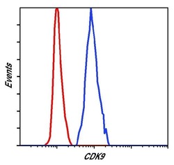
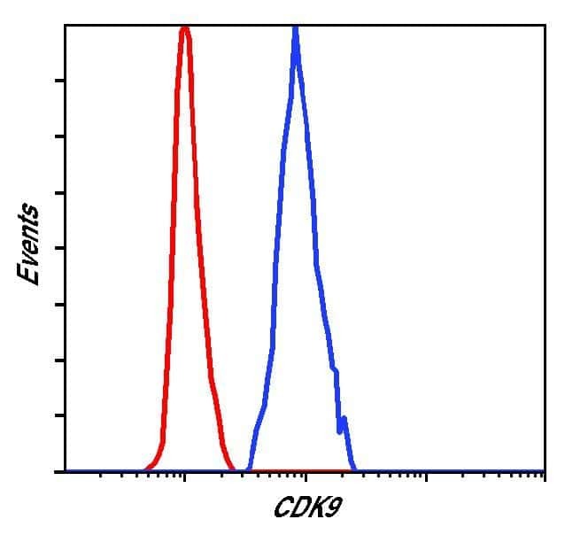
- Flow cytometric analysis of CDK9 in Jurkat cells using a CDK9 monoclonal antibody (Product # MA5-14912) (blue) compared to a nonspecific negative control antibody (red).
- Submitted by
- Invitrogen Antibodies (provider)
- Main image

- Experimental details
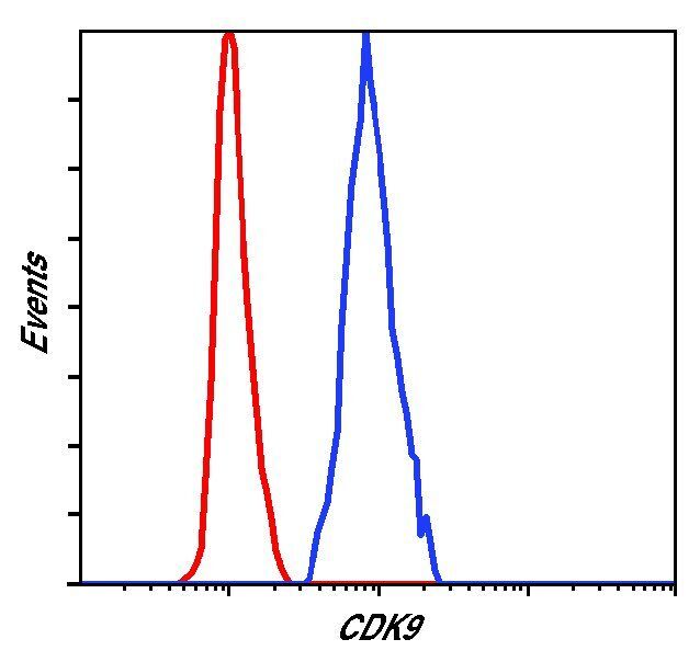
- Flow cytometric analysis of CDK9 in Jurkat cells using a CDK9 monoclonal antibody (Product # MA5-14912) (blue) compared to a nonspecific negative control antibody (red).
Supportive validation
- Submitted by
- Invitrogen Antibodies (provider)
- Main image
- Experimental details
- NULL

- Submitted by
- Invitrogen Antibodies (provider)
- Main image
- Experimental details
- Immunoprecipitation of CDK9 from HeLa cells using a CDK9 monoclonal antibody (Product # MA5-14912) followed by Western blot analysis using the same antibody. (Lane 1) 5% input.
- Submitted by
- Invitrogen Antibodies (provider)
- Main image
- Experimental details
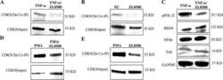
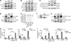
- Figure 6 ZL0580 inhibits Tat transactivation and key factors in HIV transcription elongation. ( A and B ) Western blot measurement of Tat and NF-kappaB ( A ) and cellular proteins involved in transcription elongation ( B ) in WT J-Lat cells 24 hours after treatment. ( C ) Co-IP analysis for binding of CDK9 to Tat or BRD4 in WT J-Lat cells 24 hours after treatment. Control IgG Co-IP and input CDK9 were used as controls. Total/input CDK9 blots in panels ( B and C ) represent the same experiment. ( D ) ELL2 protein expression in WT and BRD4-KO J-Lat cells 24 hours after treatment. ( E ) ELL2 mRNA expression by qPCR in WT J-Lat cells 24 hours after treatment. ( F ) Effect of protease inhibition by MG132 on ELL2 protein levels in WT J-Lat cells. Cells were pretreated with proteasome inhibitor MG-132 for 6 hours (bottom) or not treated (top), followed by treatment with PMA or PMA+ZL0580 (10 muM) for 18 hours. ELL2 protein was measured by Western blot. ( G ) Phosphorylated RNAPII (Ser 2 CTD) in WT (top) and BRD4-KO (bottom) J-Lat cells after different treatments. Loading control GAPDH in panel ( D and G ) represents the same experiment. ( H and I ) ChIP-qPCR analysis for recruitment of Tat ( H ) or BRD4 ( I ) to HIV 5' LTR in PMA-activated or unstimulated J-Lat cells 24 hours after treatment. ChIP using control IgG was included for normalization. qPCR was conducted to quantify the precipitated HIV 5' LTR region. Data were normalized to NC. Error bars ( E ,
