Explore
 Validate
 Learn
Learn Western blot
 ELISA
ELISA Immunohistochemistry
Antibody data
- Antibody Data
- Antigen structure
- References [0]
- Comments [0]
- Validations
- Immunohistochemistry [2]
Submit
Validation data
Reference
Comment
Report error
- Product number
- MA5-15849 - Provider product page

- Provider
- Invitrogen Antibodies
- Product name
- CDK9 Monoclonal Antibody (1B5A7)
- Antibody type
- Monoclonal
- Antigen
- Purifed from natural sources
- Description
- MA5-15849 targets CDK9 in indirect ELISA, IHC and WB applications and shows reactivity with Human samples. The MA5-15849 immunogen is purified recombinant fragment of human CDK9 expressed in E. Coli. . MA5-15849 detects CDK9 which has a predicted molecular weight of approximately 43kDa.
- Reactivity
- Human
- Host
- Mouse
- Isotype
- IgG
- Antibody clone number
- 1B5A7
- Vial size
- 100 μL
- Concentration
- Conc. Not Determined
- Storage
- Store at 4°C short term. For long term storage, store at -20°C, avoiding freeze/thaw cycles.
No comments: Submit comment
Supportive validation
- Submitted by
- Invitrogen Antibodies (provider)
- Main image
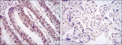
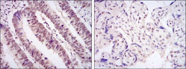
- Experimental details
- Immunohistochemical analysis of paraffin-embedded rectum cancer tissues (left) and placenta tissues (right) using CDK9 monoclonal antibody (Product # MA5-15849) followed with DAB staining.
- Submitted by
- Invitrogen Antibodies (provider)
- Main image
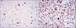
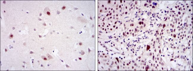
- Experimental details
- Immunohistochemical analysis of paraffin-embedded brain tissues (left) and esophageal cancer tissues (right) using CDK9 monoclonal antibody (Product # MA5-15849) followed with DAB staining.