 Explore
 Validate
 Learn
Learn Western blot
 Immunocytochemistry
Antibody data
- Antibody Data
- Antigen structure
- References [0]
- Comments [0]
- Validations
- Immunocytochemistry [1]
Submit
Validation data
Reference
Comment
Report error
- Product number
- R1184 - Provider product page

- Provider
- Acris Antibodies GmbH
- Proper citation
- Acris Antibodies GmbH Cat#R1184, RRID:AB_977046
- Product name
- anti CDK9
- Antibody type
- Polyclonal
- Antigen
- Multiple synthetic peptides corresponding to C-terminal and N-terminal domains of the protein coded by the Human gene cdk9 (PITALRE).
- Reactivity
- Human, Mouse, Rat
- Host
- Rabbit
- Vial size
- 0.1 ml
- Concentration
- 75 mg/ml (by Refractometry)
No comments: Submit comment
Supportive validation
- Submitted by
- Acris Antibodies GmbH (provider)
- Main image
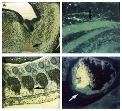
- Experimental details
- Immunocytochemical staining of Mouse tissue using CDK9 antibody (PITALRE) antiserum. The staining shows the location of mcdk9/PITALRE protein in developing Mouse tissue. Arrows indicate areas of high expression. Panel A: Peroxidase-DAB immunostaining of mcdk9/PITALRE protein in the developing Mouse brain in the differentiated region of the medulla oblongata just below the fourth ventricle. Similar staining is shown in Panel B in the dorsal root ganglia. Panel C: Fluorescein immunofluorescence of mcdk9IPITALRE in skeletal muscle. Similar staining is shown in Panel D in cardiac muscle. Other detection systems should yield similar results. Sections from each specimen were cut at 5-7 μm, mounted on glass and dried overnight at 37°C. All sections then were deparaffinized in xylene, rehydrated through a graded alcohol series and washed in phosphate-buffered saline (PBS). PBS was used for all subsequent washes and for antiserum dilution. Tissue sections were quenched sequentially in 0.5% hydrogen peroxide and blocked with diluted 10% normal goat anti-rabbit serum. Slides were incubated at 20°C for 1 h with Rabbit anti-CDK9 (1/500) dilution, washed, and then reacted with diluted Goat anti-Rabbit biotinylated antibody for 30 min. All the slides were then reacted with streptavidin-peroxidase conjugate for 30 min at 20°C. Diaminobenzidine was used as the final chromogen and hematoxylin was used as the nuclear counterstain. Negative controls for each tissue section were prepared by substituting the primary antiserum with preimmune serum.
