Explore
 Validate
 Learn
Learn Western blot
Antibody data
- Antibody Data
- Antigen structure
- References [1]
- Comments [0]
- Validations
- Western blot [1]
- Other assay [1]
Submit
Validation data
Reference
Comment
Report error
- Product number
- PA5-79423 - Provider product page

- Provider
- Invitrogen Antibodies
- Product name
- Bone SialoProtein Polyclonal Antibody
- Antibody type
- Polyclonal
- Antigen
- Synthetic peptide
- Description
- Reconstitute with 0.2 mL of distilled water to yield a concentration of 500 µg/mL.
- Reactivity
- Human, Mouse, Rat
- Host
- Rabbit
- Isotype
- IgG
- Vial size
- 100 µg
- Concentration
- 500 µg/mL
- Storage
- -20°C
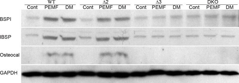
Submitted references Pulsed-electromagnetic-field induced osteoblast differentiation requires activation of genes downstream of adenosine receptors A2A and A3.
Kar NS, Ferguson D, Zhang N, Waldorff EI, Ryaby JT, DiDonato JA
PloS one 2021;16(2):e0247659
PloS one 2021;16(2):e0247659
No comments: Submit comment
Supportive validation
- Submitted by
- Invitrogen Antibodies (provider)
- Main image
- Experimental details
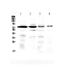
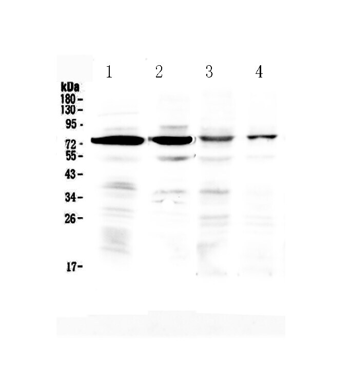
- Western blot analysis of Bone SialoProtein in Lane 1: rat brain tissue lysate, Lane 2: mouse brain tissue lysate, Lane 3: human HeLa whole cell lysate, Lane 4: human U2OS whole cell lysate using 50 µg (reducing conditions) per well. Electrophoresis was performed on 5-20% SDS-PAGE gel at 70V (Stacking gel) / 90V (Resolving gel) for 2-3 hours and protein was transferred to a nitrocellulose membrane at 150mA for 50-90 minutes. Sample was blocked with 5% Non-fat Milk/TBS for 1.5 hours at room temperature, incubated with Bone SialoProtein polyclonal antibody (Product # PA5-79423) at a dilution of 0.5 µg/mL (overnight at 4°C), followed by goat anti-rabbit IgG-HRP secondary antibody at a dilution of 1:10,000. Signal development was performed using a chemiluminescence (ECL) kit.
Supportive validation
- Submitted by
- Invitrogen Antibodies (provider)
- Main image

- Experimental details
- Fig 5 Adora3 disruption affects PEMF mediated overexpression of BSP, IBSP and Osteocalcin. Wild-type (WT), Adora2 (Delta2), Adora3 (Delta3) and Double Knock-Out (DKO) MC3T3-E1 cells were treated with PEMF (4 hours daily) or differentiation medium (DM) for 12 days and Western Blots for fractionated whole-cell proteins were probed with each of the indicated antibodies.