 Explore
 Validate
 Learn
Learn Western blot
Antibody data
- Antibody Data
- Antigen structure
- References [1]
- Comments [0]
- Validations
- Western blot [2]
- Immunohistochemistry [4]
Submit
Validation data
Reference
Comment
Report error
- Product number
- PA1-20240 - Provider product page

- Provider
- Invitrogen Antibodies
- Product name
- KIF3A Polyclonal Antibody
- Antibody type
- Polyclonal
- Antigen
- Synthetic peptide
- Description
- Recommended positive controls: PC12, rat cerebellum.
- Reactivity
- Human, Mouse, Rat
- Host
- Rabbit
- Isotype
- IgG
- Vial size
- 50 µL
- Concentration
- 5.3 mg/mL
- Storage
- Store at 4°C short term. For long term storage, store at -20°C, avoiding freeze/thaw cycles.
Submitted references KIF1-binding protein interacts with KIF3A in haploid male germ cells.
Lehti MS, Kotaja N, Sironen A
Reproduction (Cambridge, England) 2015 Sep;150(3):209-16
Reproduction (Cambridge, England) 2015 Sep;150(3):209-16
No comments: Submit comment
Supportive validation
- Submitted by
- Invitrogen Antibodies (provider)
- Main image
- Experimental details
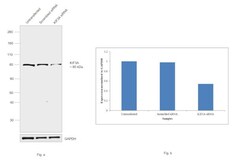
- KD of KIF3A was achieved by transfecting LNCaP with KIF3A specific siRNAs (Silencer® select Product # S21942). Western blot analysis (Fig. a) was performed using whole cell extracts from the KIF3A KD cells (Lane 3), non-specific scrambled siRNA transfected cells (Lane 2) and untransfected cells (Lane 1). The blot was probed with KIF3A Polyclonal Antibody (Product # PA1-20240, 1:1000 dilution) and Goat Anti-Rabbit IgG Secondary Antibody, HRP conjugate (Product # A27036, 1:4000 dilution). Densitometric analysis of this western blot is shown in histogram (Fig. b). Decrease in signal upon siRNA mediated knock down confirms that antibody is specific to KIF3A. .

- Submitted by
- Invitrogen Antibodies (provider)
- Main image
- Experimental details
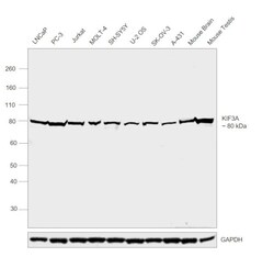
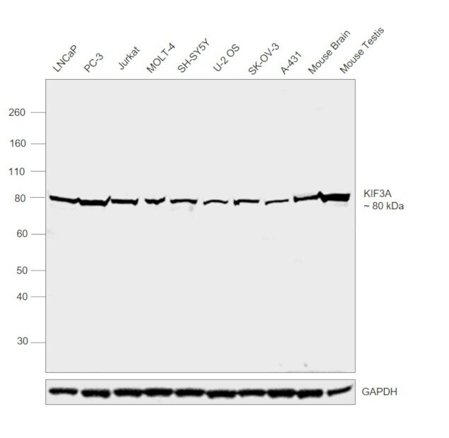
- Western blot was performed using Anti-KIF3A Polyclonal Antibody (Product # PA1-20240) and a 80 kDa band corresponding to KIF3A was observed across cell lines and tissues tested. Whole cell extracts (30 µg lysate) of LNCaP (Lane 1), PC-3 (Lane 2), Jurkat (Lane 3), MOLT-4 (Lane 4), SH-SY5Y (Lane 5), U-2 OS (Lane 6), SK-OV-3 (Lane 7), A-431 (Lane 8) and tissue extracts (30 µg lysate) of Mouse Brain (Lane 9) and Mouse Testis (Lane 10) were electrophoresed using NuPAGE™ 10% Bis-Tris Protein Gel (Product # NP0302BOX). Resolved proteins were then transferred onto a nitrocellulose membrane (Product # IB23001) by iBlot® 2 Dry Blotting System (Product # IB21001). The blot was probed with the primary antibody (1:2000 dilution) and detected by chemiluminescence Goat Anti-Rabbit IgG Secondary Antibody, HRP conjugate (Product # A27036, 1:4000 dilution) using the iBright FL 1000 (Product # A32752). Chemiluminescent detection was performed using Novex® ECL Chemiluminescent Substrate Reagent Kit (Product # WP20005)..
Supportive validation
- Submitted by
- Invitrogen Antibodies (provider)
- Main image

- Experimental details
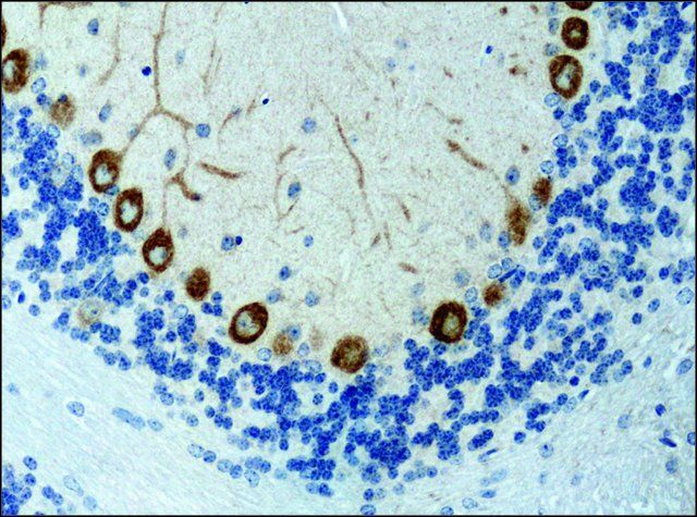
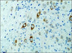
- Immunohistochemistry (Paraffin) analysis of KIF3A in rat brain tissue using KIF3A Polyclonal Antibody (Product # PA1-20240) at a dilution of 0.1 µg/mL.

- Submitted by
- Invitrogen Antibodies (provider)
- Main image

- Experimental details
- Immunohistochemistry (Paraffin) analysis of KIF3A in rat brain tissue using KIF3A Polyclonal Antibody (Product # PA1-20240) at a dilution of 0.1 µg/mL.

- Submitted by
- Invitrogen Antibodies (provider)
- Main image

- Experimental details
- Immunohistochemistry (Paraffin) analysis of KIF3A in mouse brain tissue using KIF3A Polyclonal Antibody (Product # PA1-20240) at a dilution of 0.4 µg/mL.
- Submitted by
- Invitrogen Antibodies (provider)
- Main image
- Experimental details
- Immunohistochemistry (Paraffin) analysis of KIF3A in mouse brain tissue using KIF3A Polyclonal Antibody (Product # PA1-20240) at a dilution of 0.4 µg/mL.
