Explore
 Validate
 Learn
LearnPA5-65088
antibody from Invitrogen Antibodies
Targeting: MCM4
CDC21, CDC54, hCdc21, MGC33310, P1-Cdc21
 Western blot
Antibody data
- Antibody Data
- Antigen structure
- References [0]
- Comments [0]
- Validations
- Western blot [3]
- Immunocytochemistry [2]
Submit
Validation data
Reference
Comment
Report error
- Product number
- PA5-65088 - Provider product page

- Provider
- Invitrogen Antibodies
- Product name
- MCM4 Polyclonal Antibody
- Antibody type
- Polyclonal
- Antigen
- Recombinant full-length protein
- Description
- Immunogen sequence: SSPPQMHSSAI PLDFDVSSPL TYGTPSSRVE GTPRSGVRGT PVRQRPDLGS AQKGLQVDLQ SDGAAAEDIV ASEQSLGQKL VIWGTDVNVA ACKENFQRFL QRFIDPLAKE EENVG Highest antigen sequence identity to the following orthologs - mouse 95%, rat 93%.
- Reactivity
- Human
- Host
- Rabbit
- Isotype
- IgG
- Vial size
- 100 µL
- Concentration
- 0.2 mg/mL
- Storage
- Store at 4°C short term. For long term storage, store at -20°C, avoiding freeze/thaw cycles.
No comments: Submit comment
Supportive validation
- Submitted by
- Invitrogen Antibodies (provider)
- Main image
- Experimental details
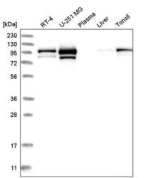
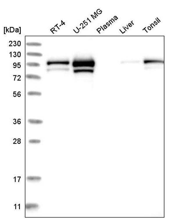
- Western blot analysis of MCM4 in human cell line RT-4, human cell line U-251 MG, human plasma, human liver tissue and human tonsil tissue. Samples were probed using a MCM4 Polyclonal Antibody (Product # PA5-65088).
- Submitted by
- Invitrogen Antibodies (provider)
- Main image

- Experimental details
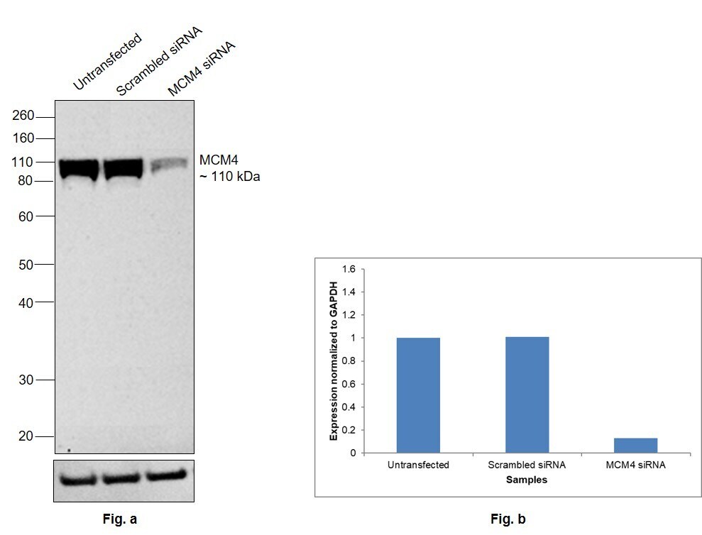
- Knockdown of MCM4 was achieved by transfecting SH-SY5Y with MCM4 specific siRNAs (Silencer® select Product # S8592, S8593). Western blot analysis (Fig. a) was performed using Nuclear enriched extracts from the MCM4 knockdown cells (lane 3), non-targeting scrambled siRNA transfected cells (lane 2) and untransfected cells (lane 1). The blot was probed with MCM4 Polyclonal Antibody (Product # PA5-65088, 1:200 dilution) and Goat anti-Rabbit IgG (H+L) Superclonal™ Recombinant Secondary Antibody, HRP (Product # A27036, 1:5000 dilution). Densitometric analysis of this western blot is shown in histogram (Fig. b). Decrease in signal upon siRNA mediated knock down confirms that antibody is specific to MCM4.
- Submitted by
- Invitrogen Antibodies (provider)
- Main image
- Experimental details
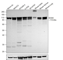
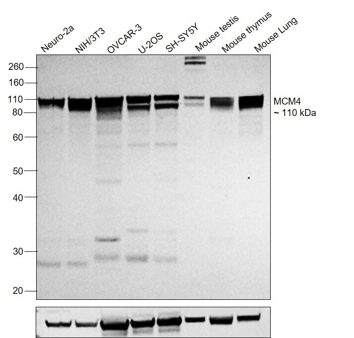
- Western blot was performed using Anti-MCM4 Polyclonal Antibody (Product # PA5-65088) and a 110 kDa band corresponding to MCM4 was observed across cell lines and tissues tested. Nuclear enriched extracts (30 µg lysate) of Neuro-2a (Lane 1), NIH/3T3 (Lane 2), NIH:OVCAR-3 (Lane 3), U-2 OS (Lane 4), SH-SY5Y (Lane 5), Mouse testis (Lane 6), Mouse Thymus (Lane 7), Mouse lung (Lane 8) were electrophoresed using NuPAGE™ 10% Bis-Tris Protein Gel (Product # NP0302BOX). Resolved proteins were then transferred onto a Nitrocellulose membrane (Product # IB23001) by iBlot® 2 Dry Blotting System (Product # IB21001). The blot was probed with the primary antibody (1:200 dilution) and detected by chemiluminescence with Goat anti-Rabbit IgG (H+L) Superclonal™ Recombinant Secondary Antibody, HRP (Product # A27036,1:5000 dilution) using the iBright FL 1000 (Product # A32752). Chemiluminescent detection was performed using Novex® ECL Chemiluminescent Substrate Reagent Kit (Product # WP20005).
Supportive validation
- Submitted by
- Invitrogen Antibodies (provider)
- Main image
- Experimental details
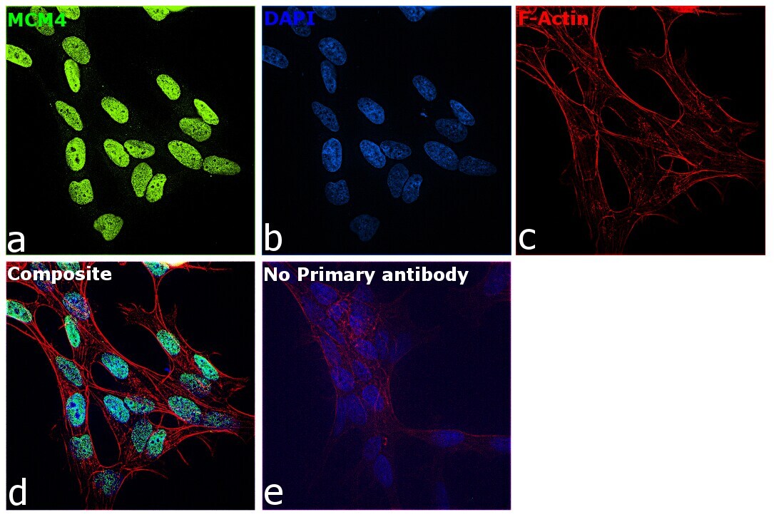
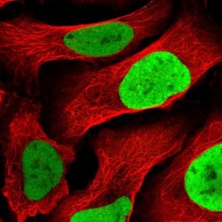
- Immunofluorescent staining of MCM4 in human cell line U-2 OS shows localization to nucleoplasm. Samples were probed using a MCM4 Polyclonal Antibody (Product # PA5-65088).

- Submitted by
- Invitrogen Antibodies (provider)
- Main image

- Experimental details
- Immunofluorescence analysis of MCM4 was performed using 70% confluent log phase SH-SY5Y cells. The cells were fixed with 4% paraformaldehyde for 10 minutes, permeabilized with 0.1% Triton™ X-100 for 15 minutes and blocked with 2% BSA for 1 hour at room temperature. The cells were labeled with MCM4 Polyclonal Antibody (Product # PA5-65088) at 1:200 dilution in 0.1% BSA and incubated overnight at 4 degree and then labeled with Goat anti-Rabbit IgG (H+L) Superclonal™ Secondary Antibody, Alexa Fluor® 488 conjugate (Product # A27034), (1:2000 dilution) for 45 minutes at room temperature (Panel a: Green). Nuclei (Panel b: Blue) were stained with ProLong™ Diamond Antifade Mountant with DAPI (Product # P36962). F-actin (Panel c: Red) was stained with Rhodamine Phalloidin (Product # R415, 1:300). Panel d represents the merged image of SH-SY5Y cells showing nuclear localization for MCM4. Panel e represents control cells with no primary antibody to assess background. The images were captured at 60X magnification.