Explore
 Validate
 Learn
Learn Western blot
 Immunocytochemistry
Antibody data
- Antibody Data
- Antigen structure
- References [5]
- Comments [0]
- Validations
- Immunocytochemistry [2]
- Other assay [4]
Submit
Validation data
Reference
Comment
Report error
- Product number
- PA3-015 - Provider product page

- Provider
- Invitrogen Antibodies
- Product name
- SEC61B Polyclonal Antibody
- Antibody type
- Polyclonal
- Antigen
- Synthetic peptide
- Description
- PA3-015 detects human, canine and porcine, and rat sec61 beta from microsomes and shows no cross-reactivity with other sec61 isoforms. PA3-015 has been successfully used in Western blot procedures. By Western blot, this antibody detects an ~14 kDa protein representing sec61 beta from pig microsomes. The PA3-015 immunogen is a synthetic peptide corresponding to residues M(1) P G P T P S G T N V G S S G R S P S K(20) of canine sec61 beta. This sequence is completely conserved between human, mouse, and canine.
- Reactivity
- Human, Mouse, Rat, Canine, Porcine
- Host
- Rabbit
- Isotype
- IgG
- Vial size
- 100 μL
- Concentration
- Conc. Not Determined
- Storage
- -20°C, Avoid Freeze/Thaw Cycles
Submitted references Genomic Action of Sigma-1 Receptor Chaperone Relates to Neuropathic Pain.
The Alzheimer's disease-associated C99 fragment of APP regulates cellular cholesterol trafficking.
Treatment of Epidermal Growth Factor (EGF) enhances Nuclear Maturation of Porcine Oocytes and Stimulates Expression of ER/Golgi Transport Proteins.
mTORC1 phosphorylates LARP6 to stimulate type I collagen expression.
Characterization of binding of LARP6 to the 5' stem-loop of collagen mRNAs: implications for synthesis of type I collagen.
Wang SM, Goguadze N, Kimura Y, Yasui Y, Pan B, Wang TY, Nakamura Y, Lin YT, Hogan QH, Wilson KL, Su TP, Wu HE
Molecular neurobiology 2021 Jun;58(6):2523-2541
Molecular neurobiology 2021 Jun;58(6):2523-2541
The Alzheimer's disease-associated C99 fragment of APP regulates cellular cholesterol trafficking.
Montesinos J, Pera M, Larrea D, Guardia-Laguarta C, Agrawal RR, Velasco KR, Yun TD, Stavrovskaya IG, Xu Y, Koo SY, Snead AM, Sproul AA, Area-Gomez E
The EMBO journal 2020 Oct 15;39(20):e103791
The EMBO journal 2020 Oct 15;39(20):e103791
Treatment of Epidermal Growth Factor (EGF) enhances Nuclear Maturation of Porcine Oocytes and Stimulates Expression of ER/Golgi Transport Proteins.
Yong H, Oh HI, Lee SH, Cheong HT, Yang BK, Park CK
Development & reproduction 2017 Jun;21(2):131-138
Development & reproduction 2017 Jun;21(2):131-138
mTORC1 phosphorylates LARP6 to stimulate type I collagen expression.
Zhang Y, Stefanovic B
Scientific reports 2017 Jan 23;7:41173
Scientific reports 2017 Jan 23;7:41173
Characterization of binding of LARP6 to the 5' stem-loop of collagen mRNAs: implications for synthesis of type I collagen.
Stefanovic L, Longo L, Zhang Y, Stefanovic B
RNA biology 2014;11(11):1386-401
RNA biology 2014;11(11):1386-401
No comments: Submit comment
Supportive validation
- Submitted by
- Invitrogen Antibodies (provider)
- Main image

- Experimental details
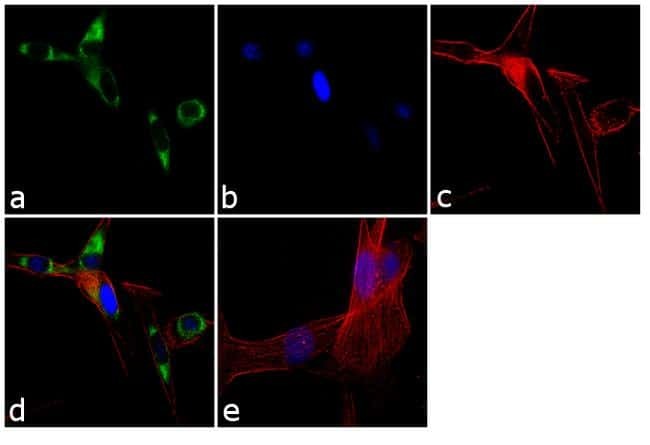
- Immunofluorescence analysis of SEC61B was performed using 70% confluent log phase MDCK cells. The cells were fixed with 4% paraformaldehyde for 10 minutes, permeabilized with 0.1% Triton™ X-100 for 10 minutes, and blocked with 1% BSA for 1 hour at room temperature. The cells were labeled with SEC61B Rabbit Polyclonal Antibody (Product # PA3-015) at 1:250 dilution in 0.1% BSA and incubated for 3 hours at room temperature and then labeled with Goat anti-Rabbit IgG (H+L) Superclonal™ Secondary Antibody, Alexa Fluor® 488 conjugate (Product # A27034) at a dilution of 1:2000 for 45 minutes at room temperature (Panel a: green). Nuclei (Panel b: blue) were stained with SlowFade® Gold Antifade Mountant with DAPI (Product # S36938). F-actin (Panel c: red) was stained with Rhodamine Phalloidin (Product # R415, 1:300). Panel d represents the merged image showing cytoplasmic localization. Panel e shows the no primary antibody control. The images were captured at 60X magnification.
- Submitted by
- Invitrogen Antibodies (provider)
- Main image

- Experimental details
- Immunofluorescence analysis of SEC61B was performed using 70% confluent log phase MDCK cells. The cells were fixed with 4% paraformaldehyde for 10 minutes, permeabilized with 0.1% Triton™ X-100 for 10 minutes, and blocked with 1% BSA for 1 hour at room temperature. The cells were labeled with SEC61B Rabbit Polyclonal Antibody (Product # PA3-015) at 1:250 dilution in 0.1% BSA and incubated for 3 hours at room temperature and then labeled with Goat anti-Rabbit IgG (Heavy Chain) Superclonal™ Secondary Antibody, Alexa Fluor® 488 conjugate (Product # A27034) at a dilution of 1:2000 for 45 minutes at room temperature (Panel a: green). Nuclei (Panel b: blue) were stained with SlowFade® Gold Antifade Mountant with DAPI (Product # S36938). F-actin (Panel c: red) was stained with Rhodamine Phalloidin (Product # R415, 1:300). Panel d represents the merged image showing cytoplasmic localization. Panel e shows the no primary antibody control. The images were captured at 60X magnification.

Supportive validation
- Submitted by
- Invitrogen Antibodies (provider)
- Main image
- Experimental details
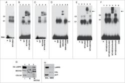
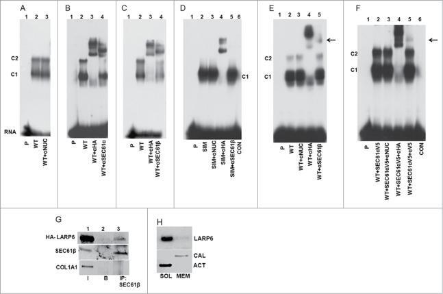
- Figure 7. Identification of SEC61 in complex with LARP6 and 5'SL. ( A ) Control supershift assay. WT HA-LARP6 was expressed in HEK293 cells and gel shift performed with cell extract without antibody (lane 2) or with anti-nucleolin antibody added (lane 3). Lane 1 is probe alone. Migration of C1 and C2 complexes is indicated. ( B ) SEC61alpha in complex with LARP6 and 5'SL. WT HA-LARP6 was expressed in HEK293 cells and gel shift performed with cell extracts without antibody (lane 2), with anti-HA antibody added (lane 3) or with anti-SEC61alpha antibody added (lane 4). Lane 1 is probe alone. ( C ) SEC61beta in complex with LARP6 and 5'SL. Experiment as in B, except anti-SEC61beta antibody was added (lane 4). ( D ) SIM mutant does not complex with SEC61. HA-tagged SIM mutant was transfected into HEK293 cells. Gel mobility shift was done with cell extracts without antibody (lane 2), with anti-nucleolin antibody added (lane 3), with anti-HA antibody added (lane 4) or with anti-SEC61beta antibody added (lane 5). Lane 1 is probe alone and lane 6 is extract of nontransfected cells. ( E ) SEC61beta in complex with LARP6 and 5'SL in extracts of human lung fibroblasts (HLFs). WT HA-LARP6 was expressed in HLFs and gel mobility shift experiment done with extract without antibody (lane 2), with anti-nucleolin antibody added (lane 3), with anti-HA antibody added (lane 4) or with anti-SEC61beta antibody added (lane 5). Lane 1 is probe alone. Arrow indicates retardation of
- Submitted by
- Invitrogen Antibodies (provider)
- Main image

- Experimental details
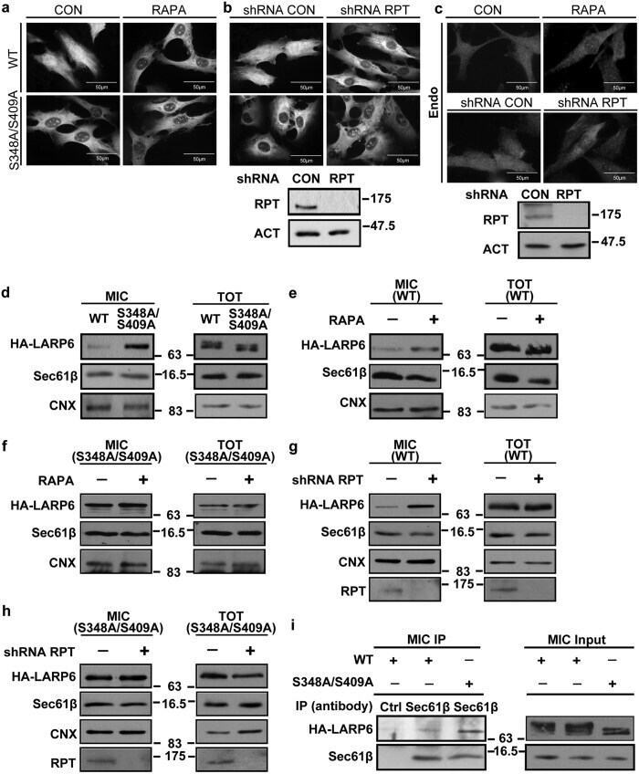
- Figure 7 Association of LARP6 with ER membrane. ( a ) Cellular localization of HA-LARP6. Wt HA-LARP6 and S348A/S409A were expressed in HLFs for 4 h and immunostained in control (CON) and rapamycin (RAPA) treated cells using anti-HA antibody. ( b ) Localization of HA-LARP6 after raptor knockdown. Raptor was knocked down by shRNA (shRPT), wt HA-LARP6 and S348A/S409A were expressed and immunostained as in ( a ). Right panel: the efficiency of raptor knockdown (RPT), loading control: beta-actin (ACT). ( c ) Cellular localization of endogenous LARP6. HLFs were treated without or with RAPA (upper panels) or transduced with lentivirus expressing control shRNA (CON) or shRNA raptor (RPT) (lower panels) and immunostained with anti-LARP6 antibody. Right panel: the efficiency of raptor knockdown (RPT). ( d ) Accumulation of S348A/S409A mutant in the microsomal fraction. Wt HA-LARP6 and S348A/S409A were expressed in HLFs and microsomal fraction (MIC, left panel) and total lysate (TOT, right panel) were analyzed by Western blot. Calnexin (CNX) and Sec61beta are shown as ER markers. ( e ) Accumulation of wt HA-LARP6 in microsomal fraction by rapamycin. Microsomal fraction and total cell lysate were analyzed as in ( d ) after treatment with rapamycin. ( f ) Microsomal accumulation of S348A/S409A mutant. Experiment as in ( e ), except S348A/S409A was analyzed. ( g ) Accumulation of wt HA-LARP6 in microsomal fraction after raptor knockdown. Microsomal fraction (MIC, left panel) and total cell
- Submitted by
- Invitrogen Antibodies (provider)
- Main image
- Experimental details
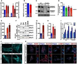
- Figure EV1 Increases in MAM -localized C99 induces cholesterol internalization and trafficking A Quantification of free cholesterol and cholesteryl ester levels in total homogenates from fibroblasts from familial (FAD) and sporadic (SAD) AD patients by lipidomics analysis. Lipid units are represented as molar mass over total moles of lipids analyzed (mol%). Graphs represent fold change over controls (WT). Unpaired t -test vs. WT ( n = 4-8; * P < 0.05, ** P < 0.01). B Quantification of HMGCR enzymatic activity showed a decreased rate of the de novo synthesis of cholesterol in fibroblasts from FAD patients. One-sample t -test vs. WT ( n = 3-4; * P < 0.05). C Measurement of SREBP2 levels by WB showed reductions in its mature/active form in DAPT-treated WT cells, and a reduction in both the full-length/precursor and mature forms in PS-DKO cells. Note how reductions in SREBP2 levels in mutant cells were abrogated upon BACE1 inhibition (BI). Coomassie staining is shown as a loading control. D, E Quantification of cholesterol uptake in (D) FAD fibroblasts and (E) neuroblastoma cell lines (Neuro-2a) where PS1, or both PS1 and PS2, had been transiently silenced. Dashed line indicates control levels. One-sample t -test vs. WT ( n = 3; * P < 0.05). F Western blot of isolated MAM and ER fractions used in Fig 1 E and F. Erlin-2 and ERp72 were used as MAM and ER markers, respectively (TH: total homogenate, CM: crude membrane). G, H Ratio of cholesteryl esters (CE):free cholesterol levels m
- Submitted by
- Invitrogen Antibodies (provider)
- Main image
- Experimental details
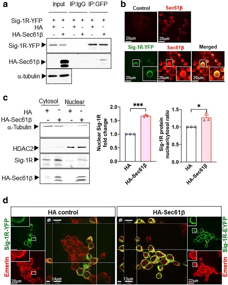
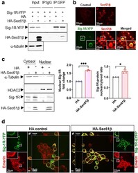
- Fig. 3 Sig-1R translocates to the nuclear envelope via Sec61beta. a Sig-1R interacts with Sec61beta. In N2a cells, HA-tagged Sec61beta and Sig-1R-EYFP were overexpressed. Immunoprecipitation with GFP antibody shows an interaction between Sig-1R and Sec61beta. b Sig-1R colocalizes with endogenous Sec61beta. Confocal images demonstrate colocalization of immunoreactive EYFP-tagged Sig-1R (green) and Sec61beta (red) in Sig-1R-EYFP overexpressing N2a cells. c Increase of Sig-1R in nuclear fraction in HA-Sec61beta-expressing N2a cells. Subcellular fractionation followed by western blotting shows an increase of Sig-1R in the nuclear fraction (left and middle panels) as reflected in the nuclear/cytosol ratio (right panel). d Colocalization of Sig-1R and emerin in Sig-1R-EYFP and HA-Sec61beta overexpressing N2a cells. Confocal images indicate perinuclear colocalization of immunoreactive EYFP-tagged Sig-1R (green) and emerin (red). Data are shown as means +- SEM; Student's t test; * P < 0.05, *** P < 0.001