Explore
 Validate
 Learn
Learn Western blot
Antibody data
- Antibody Data
- Antigen structure
- References [0]
- Comments [0]
- Validations
- Western blot [2]
- Immunocytochemistry [1]
- Flow cytometry [1]
Submit
Validation data
Reference
Comment
Report error
- Product number
- PA5-49256 - Provider product page

- Provider
- Invitrogen Antibodies
- Product name
- PRSSL1 Polyclonal Antibody
- Antibody type
- Polyclonal
- Antigen
- Synthetic peptide
- Description
- Predicted to react with mouse and rat based on sequence homology.
- Reactivity
- Human
- Host
- Rabbit
- Isotype
- IgG
- Vial size
- 400 μL
- Concentration
- 0.27 mg/mL
- Storage
- Store at 4°C short term. For long term storage, store at -20°C, avoiding freeze/thaw cycles.
No comments: Submit comment
Supportive validation
- Submitted by
- Invitrogen Antibodies (provider)
- Main image
- Experimental details
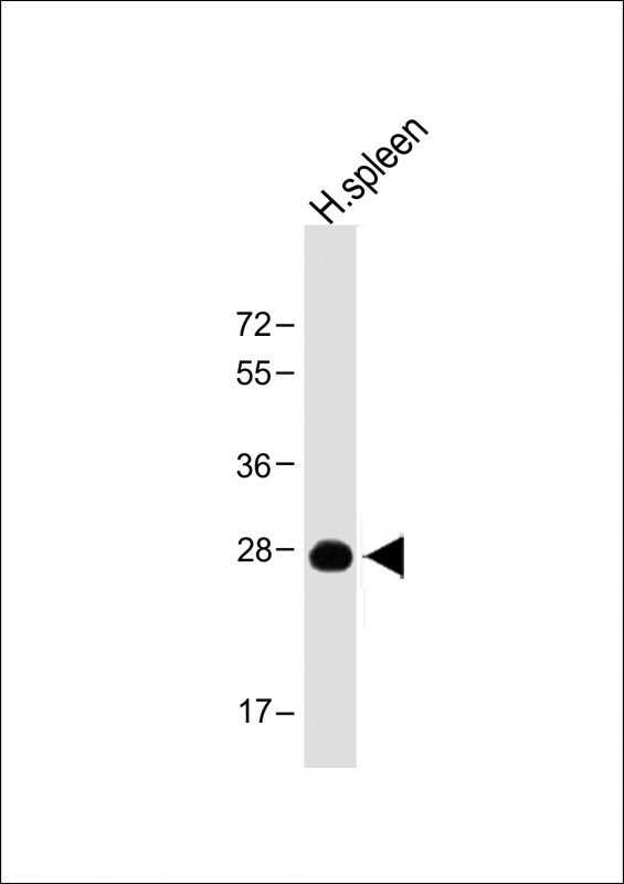
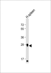
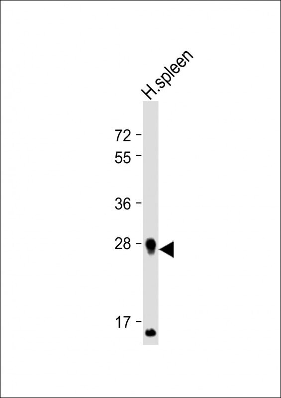
- Western blot analysis of PRSSL1 in human spleen lysate. Samples were incubated with PRSSL1 polyclonal antibody (Product # PA5-49256) using a dilution of 1:4,000 followed by Goat Anti-Rabbit IgG, (H+L), Peroxidase conjugated at a dilution of 1:10,000. Lysates/proteins: 20 µg per lane. Predicted band size: 30 kDa. Blocking/Dilution buffer: 5% NFDM/TBST.
- Submitted by
- Invitrogen Antibodies (provider)
- Main image
- Experimental details
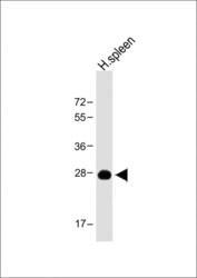
- Western blot analysis of PRSSL1 in human spleen lysate. Samples were incubated with PRSSL1 polyclonal antibody (Product # PA5-49256) using a dilution of 1:2,000 followed by Goat Anti-Rabbit IgG, (H+L), Peroxidase conjugated at a dilution of 1:10,000. Lysates/proteins: 20 µg per lane. Predicted band size: 30 kDa. Blocking/Dilution buffer: 5% NFDM/TBST.
Supportive validation
- Submitted by
- Invitrogen Antibodies (provider)
- Main image
- Experimental details
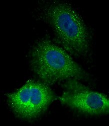
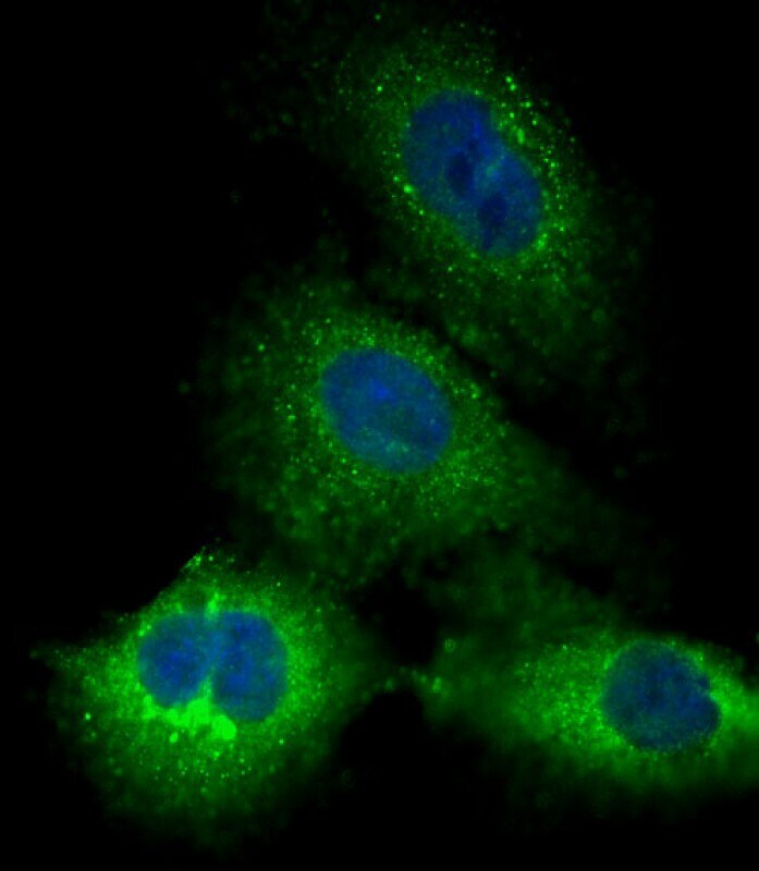
- Immunocytochemistry analysis of PRSSL1 in A549 cells. Samples were incubated with PRSSL1 polyclonal antibody (Product # PA5-49256) using a dilution of 1:25 followed by Dylight® 488-conjugated goat anti-Rabbit IgG at a dilution of 1:200 (green). Cells were 4% paraformaldehyde-fixed, 0. 1% Triton X-100 permeabilized. Immunofluorescence image showing cytoplasm staining on A549 cell line. Cytoplasmic actin is detected with Dylight® 554 Phalloidin at 1:500 dilution (red). The nuclear counter stain is DAPI (blue).
Supportive validation
- Submitted by
- Invitrogen Antibodies (provider)
- Main image

- Experimental details
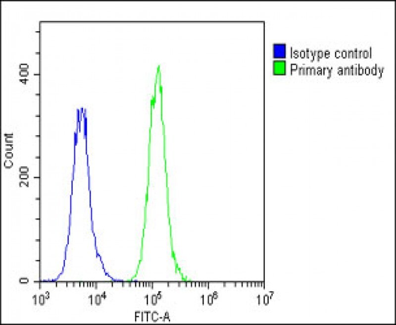
- Flow cytometry of (overlay histogram) of PRSSL1 in A549 cells (green line). Samples were incubated with PRSSL1 polyclonal antibody (Product # PA5-49256) using a dilution of 1:25 dilution for 60 min at 37°C followed by Goat-Anti-Rabbit IgG, DyLight® 488 Conjugated Highly Cross-Adsorbed at 1:200 dilution for 40 min at 37°C. The cells were fixed with 2% paraformaldehyde (10 min) and then permeabilized with 90% methanol for 10 min. The cells were then incubated in 2% bovine serum albumin to block non-specific protein-protein interactions followed by the primary antibody. Isotype control antibody (blue line) was rabbit IgG1 (1 μg/1x10^6 cells) used under the same conditions. Acquisition of >10, 000 events was performed.