Explore
 Validate
 Learn
Learn Western blot
 ELISA
ELISA Immunocytochemistry
Antibody data
- Antibody Data
- Antigen structure
- References [1]
- Comments [0]
- Validations
- Immunocytochemistry [3]
Submit
Validation data
Reference
Comment
Report error
- Product number
- 37-7400 - Provider product page

- Provider
- Invitrogen Antibodies
- Product name
- MUC5B Monoclonal Antibody (5B19-2E)
- Antibody type
- Monoclonal
- Antigen
- Synthetic peptide
- Description
- Both IHC and WB applications require the following deglycosylation protocols. Immunohistochemistry 1. Prepare the following stock solutions: a. 200 mM NaIO4 (8.556 g/200 mL water) b. Sodium-Acetate buffer, pH 4.5: Dissolve NaCl (final concentration: 0.33M) into 0.1M glacial acetic acid and adjust to pH 4.5 using 1 N NaOH. c. Sodium-thiosulfate solution: Dissolve Na2S2O3 (0.133M), NaI (0.033M), and NaHCO3 (0.033M) in water and adjust to pH 7.6 using 1M HCl. Keep solution at 4°C. 2. After deparaffinization, rehydration, and peroxidase quenching steps, mix a 1:1 solution of a and b (in step 1), add to each slide, and incubate overnight at 4°C. 3. Treat tissue with solution c for 30 minutes at 4°C. 4. Proceed with normal immunostaining protocol (blocking and antibody incubation steps). Western blotting 1. Treat transblotted membrane with 10 mM NaIO4 in 0.1M sodium acetate/5 mM EDTA, pH 5.5 overnight at 4°C in the dark. 2. After washing off the solution, incubate the membrane in a solution containing 30 mM Na2S2O3, 7.5 mM NaI, and 7.5 mM NaHCO3, pH 7.2 for 1 hour at 4°C in the dark. 3. Proceed with normal Western blot protocol (wash and blocking steps).
- Reactivity
- Human
- Host
- Mouse
- Isotype
- IgG
- Antibody clone number
- 5B19-2E
- Vial size
- 100 μg
- Concentration
- 0.5 mg/mL
- Storage
- -20°C
Submitted references Abnormalities in MUC5AC and MUC5B Protein in Airway Mucus in Asthma.
Lachowicz-Scroggins ME, Yuan S, Kerr SC, Dunican EM, Yu M, Carrington SD, Fahy JV
American journal of respiratory and critical care medicine 2016 Nov 15;194(10):1296-1299
American journal of respiratory and critical care medicine 2016 Nov 15;194(10):1296-1299
No comments: Submit comment
Supportive validation
- Submitted by
- Invitrogen Antibodies (provider)
- Main image

- Experimental details
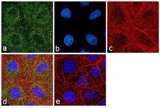
- Immunofluorescence analysis of MUC5B was performed using 70 % confluent log phase A549 cells. The cells were fixed with 4% paraformaldehyde for 10 minutes, permeabilized with 0.1% Triton X-100 for 10 minutes, and blocked with 1% BSA for 1 hour at room temperature. The cells were labeled with MUC5B (5B19-2E) Mouse Monoclonal antibody (Product # 37-7400) at 2 µg/mL in 0.1% BSA and incubated for 3 hours at room temperature and then labeled with Goat anti-Mouse IgG (H+L) Superclonal Secondary Antibody, Alexa Fluor® 488 conjugate (Product # A28175) at a dilution of 1:2000 for 45 minutes at room temperature (Panel a: green). Nuclei (Panel b: blue) were stained with SlowFade® Gold Antifade Mountant with DAPI (Product # S36938). F-actin (Panel c: red) was stained with Rhodamine Phalloidin (Product # R415, 1:300). Panel d represents the merged image showing punctated cytoplasmic localization. Panel e shows the no primary antibody control. The images were captured at 60X magnification.
- Submitted by
- Invitrogen Antibodies (provider)
- Main image
- Experimental details
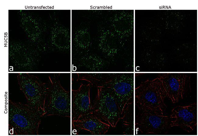
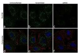
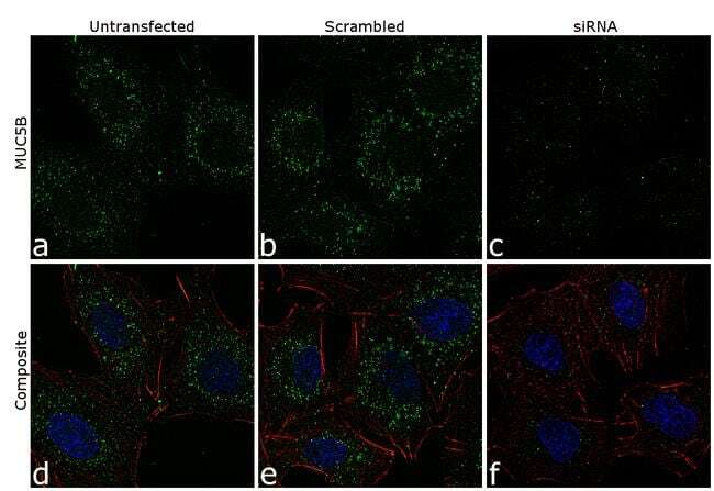
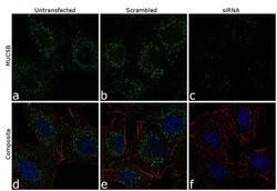
- Knockdown of MUC5B was achieved by transfecting A549 cells with MUC5B specific siRNA (Silencer® select Cat # s58031+s58030). Immunofluorescence analysis was performed on A549 cells (untransfected, panel a,d), transfected with non-specific scrambled siRNA (panels b,e) and transfected with MUC5B specific siRNA (panel c,f) Cells were fixed, permeabilized, and labelled with MUC5B Mouse monoclonal Antibody (Product # 37-7400, 5 µg/mL), followed by Goat anti-Mouse IgG (H+L) Superclonal™ Secondary Antibody, Alexa Fluor® 488 conjugate (Product # A28175, 1:2000). Nuclei (blue) were stained using SlowFade® Gold Antifade Mountant with DAPI (Product # S36938), and Rhodamine Phalloidin (Product # R415, 1:300) was used for cytoskeletal F-actin (red) staining. Loss of signal was observed upon siRNA mediated knockdown (panel c,f) confirming specificity of the antibody to MUC5B(green). The images were captured at 60X magnification.
- Submitted by
- Invitrogen Antibodies (provider)
- Main image
- Experimental details
- Knockdown of MUC5B was achieved by transfecting A549 cells with MUC5B specific siRNA (Silencer® select Cat # s58031+s58030). Immunofluorescence analysis was performed on A549 cells (untransfected, panel a,d), transfected with non-specific scrambled siRNA (panels b,e) and transfected with MUC5B specific siRNA (panel c,f) Cells were fixed, permeabilized, and labelled with MUC5B Mouse monoclonal Antibody (Product # 37-7400, 5 µg/mL), followed by Goat anti-Mouse IgG (H+L) Superclonal™ Secondary Antibody, Alexa Fluor® 488 conjugate (Product # A28175, 1:2000). Nuclei (blue) were stained using SlowFade® Gold Antifade Mountant with DAPI (Product # S36938), and Rhodamine Phalloidin (Product # R415, 1:300) was used for cytoskeletal F-actin (red) staining. Loss of signal was observed upon siRNA mediated knockdown (panel c,f) confirming specificity of the antibody to MUC5B(green). The images were captured at 60X magnification.