 Explore
 Validate
 Learn
Learn Immunocytochemistry
Antibody data
- Antibody Data
- Antigen structure
- References [33]
- Comments [0]
- Validations
- Immunocytochemistry [1]
- Immunohistochemistry [1]
Submit
Validation data
Reference
Comment
Report error
- Product number
- HPA008246 - Provider product page

- Provider
- Atlas Antibodies
- Proper citation
- Atlas Antibodies Cat#HPA008246, RRID:AB_1854203
- Product name
- Anti-MUC5B
- Antibody type
- Polyclonal
- Description
- Polyclonal Antibody against Human MUC5B, Gene description: mucin 5B, oligomeric mucus/gel-forming, Alternative Gene Names: MG1, MUC5, Validated applications: IHC, ICC, Uniprot ID: Q9HC84, Storage: Store at +4°C for short term storage. Long time storage is recommended at -20°C.
- Reactivity
- Human
- Host
- Rabbit
- Conjugate
- Unconjugated
- Isotype
- IgG
- Vial size
- 100 µl
- Concentration
- 0.2 mg/ml
- Storage
- Store at +4°C for short term storage. Long time storage is recommended at -20°C.
- Handling
- The antibody solution should be gently mixed before use.
Submitted references Isolation of Membrane Bound Mucins from Human Bronchial Epithelial Cells
Tolerance to Haemophilus influenzae infection in human epithelial cells: Insights from a primary cell-based model
Sex-dependent regulation of mucin gene transcription and airway secretion and mechanics following intra-airway IL-13 in mice with conditional loss of club cell Creb1
Transgenic ferret models define pulmonary ionocyte diversity and function
Genomic characterization and therapeutic utilization of IL-13-responsive sequences in asthma
A high-throughput cigarette smoke-treated bronchosphere model for disease-relevant phenotypic compound screening
Prime editing-mediated correction of the CFTR W1282X mutation in iPSCs and derived airway epithelial cells
Immunohistochemical detection of MUC5AC and MUC5B mucins in ferrets
Club cell CREB regulates the goblet cell transcriptional network and pro-mucin effects of IL-1B
Human intestinal organoids from Cronkhite-Canada syndrome patients reveal link between serotonin and proliferation
RSV-induced expanded ciliated cells contribute to bronchial wall thickening.
Elevated Protein Kinase A Activity in Stomach Mesenchyme Disrupts Mesenchymal-epithelial Crosstalk and Induces Preneoplasia
A Proximal-to-Distal Survey of Healthy Adult Human Small Intestine and Colon Epithelium by Single-Cell Transcriptomics
Bronchial epithelium epithelial-mesenchymal plasticity forms aberrant basaloid-like cells in vitro.
Goblet Cell Hyperplasia Increases SARS-CoV-2 Infection in Chronic Obstructive Pulmonary Disease.
Overexpression of Substance P in pig airways increases MUC5AC through an NF‐kβ pathway
Transcriptional analysis of cystic fibrosis airways at single-cell resolution reveals altered epithelial cell states and composition
Pharmacological targeting of host chaperones protects from pertussis toxin in vitro and in vivo
SOX21 modulates SOX2-initiated differentiation of epithelial cells in the extrapulmonary airways
Susceptibility of primary human airway epithelial cells to Bordetella pertussis adenylate cyclase toxin in two- and three-dimensional culture conditions
Investigation on Ciliary Functionality of Different Airway Epithelial Cell Lines in Three-Dimensional Cell Culture
Single-Cell Reconstruction of Human Basal Cell Diversity in Normal and Idiopathic Pulmonary Fibrosis Lungs
In vitro 3D culture lung model from expanded primary cystic fibrosis human airway cells
Host–Pathogen Responses to Pandemic Influenza H1N1pdm09 in a Human Respiratory Airway Model
Human Nasal Epithelial Organoids for Therapeutic Development in Cystic Fibrosis
Optimization of Normal Human Bronchial Epithelial (NHBE) Cell 3D Cultures for in vitro Lung Model Studies
Characterizing smoking-induced transcriptional heterogeneity in the human bronchial epithelium at single-cell resolution
Characterization of an immortalized human small airway basal stem/progenitor cell line with airway region-specific differentiation capacity
Expansion of airway basal epithelial cells from primary human non‐small cell lung cancer tumors
Surface modification of a POSS-nanocomposite material to enhance cellular integration of a synthetic bioscaffold
Loss of Cystic Fibrosis Transmembrane Conductance Regulator Function Produces Abnormalities in Tracheal Development in Neonatal Pigs and Young Children
Tissue-Specific Protein Expression in Human Cells, Tissues and Organs
Carpenter J, Kesimer M
2024;2763
2024;2763
Tolerance to Haemophilus influenzae infection in human epithelial cells: Insights from a primary cell-based model
Guillard T, Kappler U, Henningham A, Nasreen M, Yamamoto A, Buultjens A, Stinear T, Sly P, Fantino E
PLOS Pathogens 2024;20(7):e1012282
PLOS Pathogens 2024;20(7):e1012282
Sex-dependent regulation of mucin gene transcription and airway secretion and mechanics following intra-airway IL-13 in mice with conditional loss of club cell Creb1
Sponchiado M, Fagan A, Mata L, Bonilla A, Trevizan-Baú P, Prabhakaran S, Reznikov L
Frontiers in Physiology 2024;15
Frontiers in Physiology 2024;15
Kanke K, Rayner R, Abel E, Venugopalan A, Suu M, Stack J, Nouri R, Guo G, Vetter T, Cormet-Boyaka E, Hester M, Vaidyanathan S
2024
2024
Transgenic ferret models define pulmonary ionocyte diversity and function
Yuan F, Gasser G, Lemire E, Montoro D, Jagadeesh K, Zhang Y, Duan Y, Ievlev V, Wells K, Rotti P, Shahin W, Winter M, Rosen B, Evans I, Cai Q, Yu M, Walsh S, Acevedo M, Pandya D, Akurathi V, Dick D, Wadas T, Joo N, Wine J, Birket S, Fernandez C, Leung H, Tearney G, Verkman A, Haggie P, Scott K, Bartels D, Meyerholz D, Rowe S, Liu X, Yan Z, Haber A, Sun X, Engelhardt J
Nature 2023;621(7980):857-867
Nature 2023;621(7980):857-867
Genomic characterization and therapeutic utilization of IL-13-responsive sequences in asthma
Koh K, Bonser L, Eckalbar W, Yizhar-Barnea O, Shen J, Zeng X, Hargett K, Sun D, Zlock L, Finkbeiner W, Ahituv N, Erle D
Cell Genomics 2023;3(1):100229
Cell Genomics 2023;3(1):100229
A high-throughput cigarette smoke-treated bronchosphere model for disease-relevant phenotypic compound screening
Singanayagam A, Beri P, Woo Y, Schierenbeck K, Chen K, Barnes S, Ross O, Krutil D, Quackenbush D, Fang B, Walker J, Barnes W, Toyama E
PLOS ONE 2023;18(6):e0287809
PLOS ONE 2023;18(6):e0287809
Prime editing-mediated correction of the CFTR W1282X mutation in iPSCs and derived airway epithelial cells
Branchini A, Li C, Liu Z, Anderson J, Liu Z, Tang L, Li Y, Peng N, Chen J, Liu X, Fu L, Townes T, Rowe S, Bedwell D, Guimbellot J, Zhao R
PLOS ONE 2023;18(11):e0295009
PLOS ONE 2023;18(11):e0295009
Immunohistochemical detection of MUC5AC and MUC5B mucins in ferrets
Meyerholz D, Leidinger M, Adam Goeken J, Businga T, Vizuett S, Akers A, Evans I, Zhang Y, Engelhardt J
BMC Research Notes 2023;16(1)
BMC Research Notes 2023;16(1)
Club cell CREB regulates the goblet cell transcriptional network and pro-mucin effects of IL-1B
Sponchiado M, Bonilla A, Mata L, Jasso-Johnson K, Liao Y, Fagan A, Moncada V, Reznikov L
Frontiers in Physiology 2023;14
Frontiers in Physiology 2023;14
Human intestinal organoids from Cronkhite-Canada syndrome patients reveal link between serotonin and proliferation
Poplaski V, Bomidi C, Kambal A, Nguyen-Phuc H, Di Rienzi S, Danhof H, Zeng X, Feagins L, Deng N, Vilar E, McAllister F, Coarfa C, Min S, Kim H, Shukla R, Britton R, Estes M, Blutt S
Journal of Clinical Investigation 2023;133(21)
Journal of Clinical Investigation 2023;133(21)
RSV-induced expanded ciliated cells contribute to bronchial wall thickening.
Talukdar SN, Osan J, Ryan K, Grove B, Perley D, Kumar BD, Yang S, Dallman S, Hollingsworth L, Bailey KL, Mehedi M
Virus research 2023 Apr 2;327:199060
Virus research 2023 Apr 2;327:199060
Elevated Protein Kinase A Activity in Stomach Mesenchyme Disrupts Mesenchymal-epithelial Crosstalk and Induces Preneoplasia
Puri P, Grimmett G, Faraj R, Gibson L, Gilbreath E, Yoder B
Cellular and Molecular Gastroenterology and Hepatology 2022;14(3):643-668.e1
Cellular and Molecular Gastroenterology and Hepatology 2022;14(3):643-668.e1
A Proximal-to-Distal Survey of Healthy Adult Human Small Intestine and Colon Epithelium by Single-Cell Transcriptomics
Burclaff J, Bliton R, Breau K, Ok M, Gomez-Martinez I, Ranek J, Bhatt A, Purvis J, Woosley J, Magness S
Cellular and Molecular Gastroenterology and Hepatology 2022;13(5):1554-1589
Cellular and Molecular Gastroenterology and Hepatology 2022;13(5):1554-1589
Bronchial epithelium epithelial-mesenchymal plasticity forms aberrant basaloid-like cells in vitro.
Uthaya Kumar DB, Motakis E, Yurieva M, Kohar V, Martinek J, Wu TC, Khoury J, Grassmann J, Lu M, Palucka K, Kaminski N, Koff JL, Williams A
American journal of physiology. Lung cellular and molecular physiology 2022 Jun 1;322(6):L822-L841
American journal of physiology. Lung cellular and molecular physiology 2022 Jun 1;322(6):L822-L841
Goblet Cell Hyperplasia Increases SARS-CoV-2 Infection in Chronic Obstructive Pulmonary Disease.
Osan J, Talukdar SN, Feldmann F, DeMontigny BA, Jerome K, Bailey KL, Feldmann H, Mehedi M
Microbiology spectrum 2022 Aug 31;10(4):e0045922
Microbiology spectrum 2022 Aug 31;10(4):e0045922
Overexpression of Substance P in pig airways increases MUC5AC through an NF‐kβ pathway
Sponchiado M, Liao Y, Atanasova K, Collins E, Schurmann V, Bravo L, Reznikov L
Physiological Reports 2021;9(3)
Physiological Reports 2021;9(3)
Transcriptional analysis of cystic fibrosis airways at single-cell resolution reveals altered epithelial cell states and composition
Carraro G, Langerman J, Sabri S, Lorenzana Z, Purkayastha A, Zhang G, Konda B, Aros C, Calvert B, Szymaniak A, Wilson E, Mulligan M, Bhatt P, Lu J, Vijayaraj P, Yao C, Shia D, Lund A, Israely E, Rickabaugh T, Ernst J, Mense M, Randell S, Vladar E, Ryan A, Plath K, Mahoney J, Stripp B, Gomperts B
Nature Medicine 2021;27(5):806-814
Nature Medicine 2021;27(5):806-814
Pharmacological targeting of host chaperones protects from pertussis toxin in vitro and in vivo
Ernst K, Mittler A, Winkelmann V, Kling C, Eberhardt N, Anastasia A, Sonnabend M, Lochbaum R, Wirsching J, Sakari M, Pulliainen A, Skerry C, Carbonetti N, Frick M, Barth H
Scientific Reports 2021;11(1)
Scientific Reports 2021;11(1)
SOX21 modulates SOX2-initiated differentiation of epithelial cells in the extrapulmonary airways
Eenjes E, Buscop-van Kempen M, Boerema-de Munck A, Edel G, Benthem F, de Kreij-de Bruin L, Schnater M, Tibboel D, Collins J, Rottier R
eLife 2021;10
eLife 2021;10
Susceptibility of primary human airway epithelial cells to Bordetella pertussis adenylate cyclase toxin in two- and three-dimensional culture conditions
Bianchi M, Sivarajan R, Walles T, Hackenberg S, Steinke M
Innate Immunity 2020;27(1):89-98
Innate Immunity 2020;27(1):89-98
Investigation on Ciliary Functionality of Different Airway Epithelial Cell Lines in Three-Dimensional Cell Culture
Lodes N, Seidensticker K, Perniss A, Nietzer S, Oberwinkler H, May T, Walles T, Hebestreit H, Hackenberg S, Steinke M
Tissue Engineering Part A 2020;26(7-8):432-440
Tissue Engineering Part A 2020;26(7-8):432-440
Single-Cell Reconstruction of Human Basal Cell Diversity in Normal and Idiopathic Pulmonary Fibrosis Lungs
Carraro G, Mulay A, Yao C, Mizuno T, Konda B, Petrov M, Lafkas D, Arron J, Hogaboam C, Chen P, Jiang D, Noble P, Randell S, McQualter J, Stripp B
American Journal of Respiratory and Critical Care Medicine 2020;202(11):1540-1550
American Journal of Respiratory and Critical Care Medicine 2020;202(11):1540-1550
In vitro 3D culture lung model from expanded primary cystic fibrosis human airway cells
Rayner R, Wellmerling J, Osman W, Honesty S, Alfaro M, Peeples M, Cormet-Boyaka E
Journal of Cystic Fibrosis 2020;19(5):752-761
Journal of Cystic Fibrosis 2020;19(5):752-761
Host–Pathogen Responses to Pandemic Influenza H1N1pdm09 in a Human Respiratory Airway Model
Pharo E, Williams S, Boyd V, Sundaramoorthy V, Durr P, Baker M
Viruses 2020;12(6):679
Viruses 2020;12(6):679
Human Nasal Epithelial Organoids for Therapeutic Development in Cystic Fibrosis
Liu Z, Anderson J, Deng L, Mackay S, Bailey J, Kersh L, Rowe S, Guimbellot J
Genes 2020;11(6):603
Genes 2020;11(6):603
Optimization of Normal Human Bronchial Epithelial (NHBE) Cell 3D Cultures for in vitro Lung Model Studies
Rayner R, Makena P, Prasad G, Cormet-Boyaka E
Scientific Reports 2019;9(1)
Scientific Reports 2019;9(1)
Characterizing smoking-induced transcriptional heterogeneity in the human bronchial epithelium at single-cell resolution
Duclos G, Teixeira V, Autissier P, Gesthalter Y, Reinders-Luinge M, Terrano R, Dumas Y, Liu G, Mazzilli S, Brandsma C, van den Berge M, Janes S, Timens W, Lenburg M, Spira A, Campbell J, Beane J
Science Advances 2019;5(12)
Science Advances 2019;5(12)
Characterization of an immortalized human small airway basal stem/progenitor cell line with airway region-specific differentiation capacity
Wang G, Lou H, Salit J, Leopold P, Driscoll S, Schymeinsky J, Quast K, Visvanathan S, Fine J, Thomas M, Crystal R
Respiratory Research 2019;20(1)
Respiratory Research 2019;20(1)
Expansion of airway basal epithelial cells from primary human non‐small cell lung cancer tumors
Hynds R, Ben Aissa A, Gowers K, Watkins T, Bosshard‐Carter L, Rowan A, Veeriah S, Wilson G, Quezada S, Swanton C, Janes S
International Journal of Cancer 2018;143(1):160-166
International Journal of Cancer 2018;143(1):160-166
Surface modification of a POSS-nanocomposite material to enhance cellular integration of a synthetic bioscaffold
Crowley C, Klanrit P, Butler C, Varanou A, Platé M, Hynds R, Chambers R, Seifalian A, Birchall M, Janes S
Biomaterials 2016;83
Biomaterials 2016;83
Loss of Cystic Fibrosis Transmembrane Conductance Regulator Function Produces Abnormalities in Tracheal Development in Neonatal Pigs and Young Children
Meyerholz D, Stoltz D, Namati E, Ramachandran S, Pezzulo A, Smith A, Rector M, Suter M, Kao S, McLennan G, Tearney G, Zabner J, McCray P, Welsh M
American Journal of Respiratory and Critical Care Medicine 2010;182(10):1251-1261
American Journal of Respiratory and Critical Care Medicine 2010;182(10):1251-1261
Tissue-Specific Protein Expression in Human Cells, Tissues and Organs
Per Oksvold M
Journal of Proteomics & Bioinformatics 2010;03(10)
Journal of Proteomics & Bioinformatics 2010;03(10)
No comments: Submit comment
Supportive validation
- Submitted by
- Atlas Antibodies (provider)
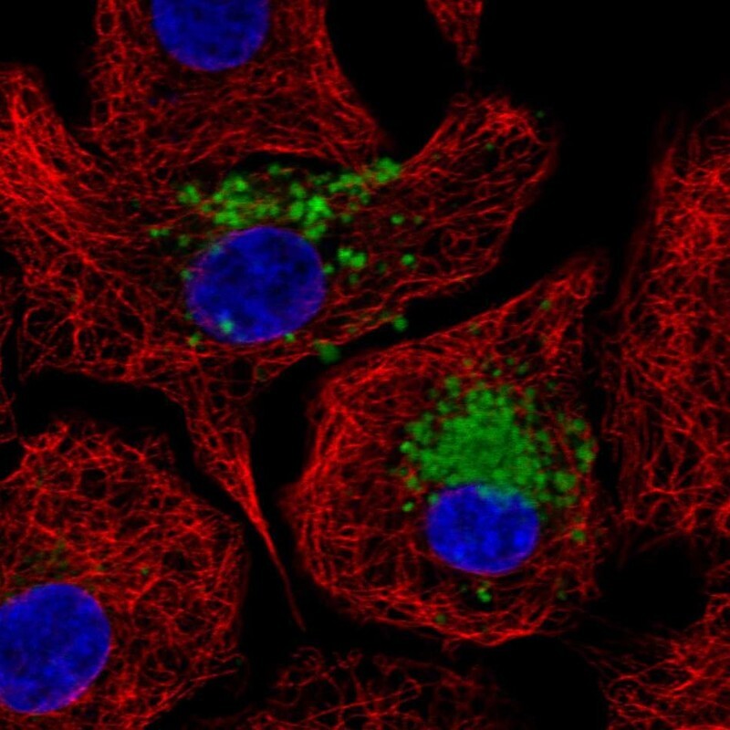
- Main image
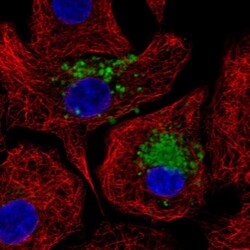
- Experimental details
- Immunofluorescent staining of human cell line A549 shows localization to vesicles.
- Sample type
- Human
Supportive validation
- Submitted by
- Atlas Antibodies (provider)
- Enhanced method
- Orthogonal validation
- Main image
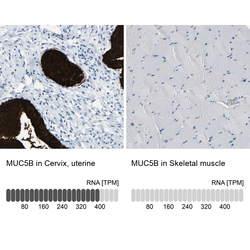
- Experimental details
- Immunohistochemistry analysis in human cervix, uterine and skeletal muscle tissues using HPA008246 antibody. Corresponding MUC5B RNA-seq data are presented for the same tissues.
- Sample type
- Human
- Protocol
- Protocol
