Explore
 Validate
 Learn
Learn Western blot
 Immunocytochemistry
Antibody data
- Antibody Data
- Antigen structure
- References [3]
- Comments [0]
- Validations
- Immunocytochemistry [1]
- Immunohistochemistry [1]
Submit
Validation data
Reference
Comment
Report error
- Product number
- HPA021191 - Provider product page

- Provider
- Atlas Antibodies
- Proper citation
- Atlas Antibodies Cat#HPA021191, RRID:AB_1856395
- Product name
- Anti-CROCC
- Antibody type
- Polyclonal
- Description
- Polyclonal Antibody against Human CROCC, Gene description: ciliary rootlet coiled-coil, rootletin, Alternative Gene Names: ROLT, rootletin, Validated applications: IHC, ICC, WB, Uniprot ID: Q5TZA2, Storage: Store at +4°C for short term storage. Long time storage is recommended at -20°C.
- Reactivity
- Human
- Host
- Rabbit
- Conjugate
- Unconjugated
- Isotype
- IgG
- Vial size
- 100 µl
- Concentration
- 0.2 mg/ml
- Storage
- Store at +4°C for short term storage. Long time storage is recommended at -20°C.
- Handling
- The antibody solution should be gently mixed before use.
Submitted references The intercentriolar fibers function as docking sites of centriolar satellites for cilia assembly
Mutations in CCNO result in congenital mucociliary clearance disorder with reduced generation of multiple motile cilia
DNA damage-induced centrosome amplification occurs via excessive formation of centriolar satellites
Ryu S, Ko D, Shin B, Rhee K
Journal of Cell Biology 2024;223(4)
Journal of Cell Biology 2024;223(4)
Mutations in CCNO result in congenital mucociliary clearance disorder with reduced generation of multiple motile cilia
Wallmeier J, Al-Mutairi D, Chen C, Loges N, Pennekamp P, Menchen T, Ma L, Shamseldin H, Olbrich H, Dougherty G, Werner C, Alsabah B, Köhler G, Jaspers M, Boon M, Griese M, Schmitt-Grohé S, Zimmermann T, Koerner-Rettberg C, Horak E, Kintner C, Alkuraya F, Omran H
Nature Genetics 2014;46(6):646-651
Nature Genetics 2014;46(6):646-651
DNA damage-induced centrosome amplification occurs via excessive formation of centriolar satellites
Löffler H, Fechter A, Liu F, Poppelreuther S, Krämer A
Oncogene 2012;32(24):2963-2972
Oncogene 2012;32(24):2963-2972
No comments: Submit comment
Supportive validation
- Submitted by
- Atlas Antibodies (provider)
- Main image
- Experimental details
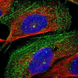
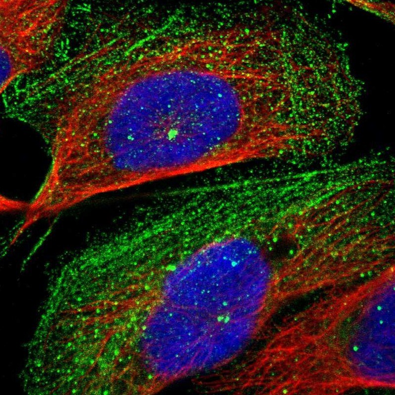
- Immunofluorescent staining of human cell line U-2 OS shows localization to plasma membrane, centrosome & actin filaments.
- Sample type
- Human
Supportive validation
- Submitted by
- Atlas Antibodies (provider)
- Enhanced method
- Orthogonal validation
- Main image
- Experimental details
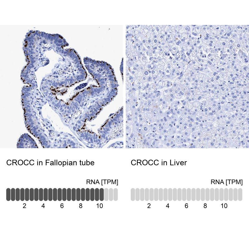
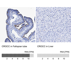
- Immunohistochemistry analysis in human fallopian tube and liver tissues using HPA021191 antibody. Corresponding CROCC RNA-seq data are presented for the same tissues.
- Sample type
- Human
- Protocol
- Protocol