Explore
 Validate
 Learn
Learn Western blot
 Immunocytochemistry
Antibody data
- Antibody Data
- Antigen structure
- References [5]
- Comments [0]
- Validations
- Western blot [1]
- Immunohistochemistry [12]
Submit
Validation data
Reference
Comment
Report error
- Product number
- NBP1-80820 - Provider product page

- Provider
- Novus Biologicals
- Proper citation
- Novus Cat#NBP1-80820, RRID:AB_11019491
- Product name
- Rabbit Polyclonal CROCC Antibody
- Antibody type
- Polyclonal
- Description
- Immunogen affinity purified. Specificity of human CROCC antibody verified on a Protein Array containing target protein plus 383 other non-specific proteins.
- Reactivity
- Human
- Host
- Rabbit
- Isotype
- IgG
- Vial size
- 0.1 ml
- Storage
- Store at 4C short term. Aliquot and store at -20C long term. Avoid freeze-thaw cycles.
Submitted references Stable centrosomal roots disentangle to allow interphase centriole independence.
Centriole splitting caused by loss of the centrosomal linker protein C-NAP1 reduces centriolar satellite density and impedes centrosome amplification.
Mutations in CCNO result in congenital mucociliary clearance disorder with reduced generation of multiple motile cilia.
DNA damage-induced centrosome amplification occurs via excessive formation of centriolar satellites.
Chemically diverse microtubule stabilizing agents initiate distinct mitotic defects and dysregulated expression of key mitotic kinases.
Mahen R
PLoS biology 2018 Apr;16(4):e2003998
PLoS biology 2018 Apr;16(4):e2003998
Centriole splitting caused by loss of the centrosomal linker protein C-NAP1 reduces centriolar satellite density and impedes centrosome amplification.
Flanagan AM, Stavenschi E, Basavaraju S, Gaboriau D, Hoey DA, Morrison CG
Molecular biology of the cell 2017 Mar 15;28(6):736-745
Molecular biology of the cell 2017 Mar 15;28(6):736-745
Mutations in CCNO result in congenital mucociliary clearance disorder with reduced generation of multiple motile cilia.
Wallmeier J, Al-Mutairi DA, Chen CT, Loges NT, Pennekamp P, Menchen T, Ma L, Shamseldin HE, Olbrich H, Dougherty GW, Werner C, Alsabah BH, Köhler G, Jaspers M, Boon M, Griese M, Schmitt-Grohé S, Zimmermann T, Koerner-Rettberg C, Horak E, Kintner C, Alkuraya FS, Omran H
Nature genetics 2014 Jun;46(6):646-51
Nature genetics 2014 Jun;46(6):646-51
DNA damage-induced centrosome amplification occurs via excessive formation of centriolar satellites.
Löffler H, Fechter A, Liu FY, Poppelreuther S, Krämer A
Oncogene 2013 Jun 13;32(24):2963-72
Oncogene 2013 Jun 13;32(24):2963-72
Chemically diverse microtubule stabilizing agents initiate distinct mitotic defects and dysregulated expression of key mitotic kinases.
Rohena CC, Peng J, Johnson TA, Crews P, Mooberry SL
Biochemical pharmacology 2013 Apr 15;85(8):1104-14
Biochemical pharmacology 2013 Apr 15;85(8):1104-14
No comments: Submit comment
Supportive validation
- Submitted by
- Novus Biologicals (provider)
- Main image
- Experimental details
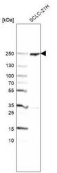
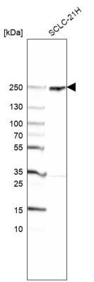
- Western Blot: CROCC Antibody [NBP1-80820] - Analysis in human cell line SCLC-21H.
Supportive validation
- Submitted by
- Novus Biologicals (provider)
- Main image
- Experimental details
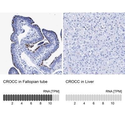
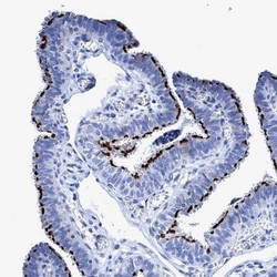
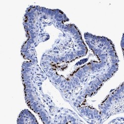
- Immunohistochemistry-Paraffin: CROCC Antibody [NBP1-80820] - Staining of human fallopian tube shows strong membranous positivity in ciliated cells.

- Submitted by
- Novus Biologicals (provider)
- Main image
- Experimental details
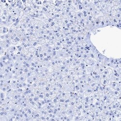
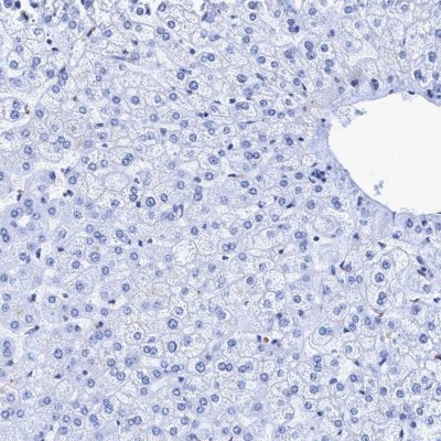
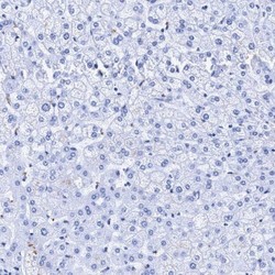
- Immunohistochemistry-Paraffin: CROCC Antibody [NBP1-80820] - Staining of human liver shows low expression as expected.
- Submitted by
- Novus Biologicals (provider)
- Main image
- Experimental details
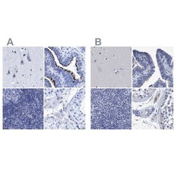
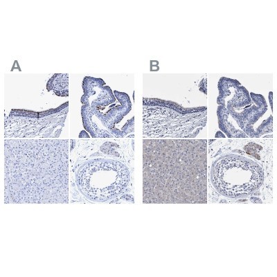
- Immunohistochemistry-Paraffin: CROCC Antibody [NBP1-80820] - Staining of human cerebral cortex, fallopian tube, lymph node and testis using Anti-CROCC antibody NBP1-80820 (A) shows similar protein distribution across tissues to independent antibody NBP1-80821 (B).
- Submitted by
- Novus Biologicals (provider)
- Main image
- Experimental details
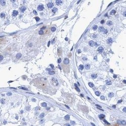
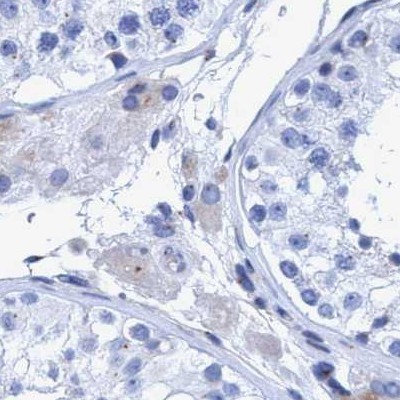
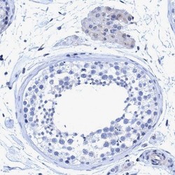
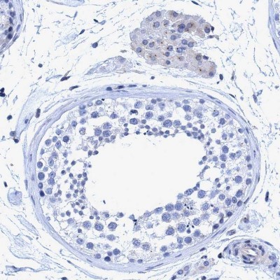
- Immunohistochemistry-Paraffin: CROCC Antibody [NBP1-80820] - Staining of human testis.
- Submitted by
- Novus Biologicals (provider)
- Main image
- Experimental details
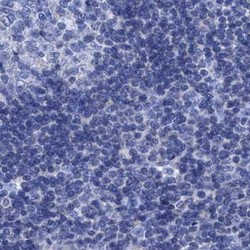
- Immunohistochemistry-Paraffin: CROCC Antibody [NBP1-80820] - Staining of human lymph node.

- Submitted by
- Novus Biologicals (provider)
- Main image
- Experimental details
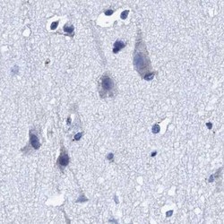
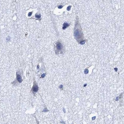
- Immunohistochemistry-Paraffin: CROCC Antibody [NBP1-80820] - Staining of human cerebral cortex.
- Submitted by
- Novus Biologicals (provider)
- Main image

- Experimental details
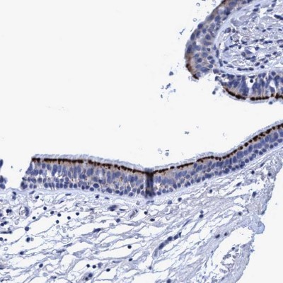
- Immunohistochemistry-Paraffin: CROCC Antibody [NBP1-80820] - Staining of human bronchus shows strong positivity in apical membrane in respiratory epithelial cells.
- Submitted by
- Novus Biologicals (provider)
- Main image

- Experimental details
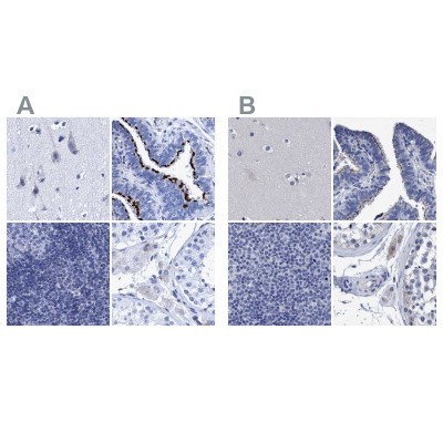
- Immunohistochemistry-Paraffin: CROCC Antibody [NBP1-80820] - Staining of human bronchus, fallopian tube, liver and testis using Anti-CROCC antibody NBP1-80820 (A) shows similar protein distribution across tissues to independent antibody NBP1-80821 (B).
- Submitted by
- Novus Biologicals (provider)
- Main image
- Experimental details
- Immunohistochemistry-Paraffin: CROCC Antibody [NBP1-80820] - Staining of human Fallopian tube shows strong membranous positivity in glandular cells.

- Submitted by
- Novus Biologicals (provider)
- Main image
- Experimental details
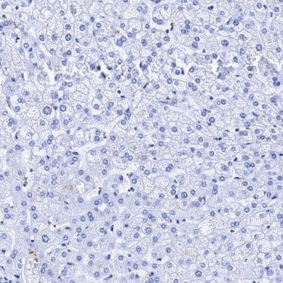
- Immunohistochemistry-Paraffin: CROCC Antibody [NBP1-80820] - Staining of human liver shows no positivity in hepatocytes as expected.
- Submitted by
- Novus Biologicals (provider)
- Main image
- Experimental details
- Immunohistochemistry-Paraffin: CROCC Antibody [NBP1-80820] - Staining of human testis shows very weak cytoplasmic positivity in Leydig cells.
- Submitted by
- Novus Biologicals (provider)
- Main image

- Experimental details
- Immunohistochemistry-Paraffin: CROCC Antibody [NBP1-80820] - Staining in human fallopian tube and liver tissues using NBP1-80820 antibody. Corresponding CROCC RNA-seq data are presented for the same tissues.