Explore
 Validate
 Learn
Learn Western blot
Antibody data
- Antibody Data
- Antigen structure
- References [0]
- Comments [0]
- Validations
- Western blot [2]
- Immunohistochemistry [1]
Submit
Validation data
Reference
Comment
Report error
- Product number
- NBP2-30080 - Provider product page

- Provider
- Novus Biologicals
- Product name
- Rabbit Polyclonal PRKD3/nPKC nu Antibody
- Antibody type
- Polyclonal
- Description
- Ammonium sulfate precipitation.
- Reactivity
- Human
- Host
- Rabbit
- Isotype
- IgG
- Vial size
- 0.4 ml
- Storage
- Store at 4C short term. Aliquot and store at -20C long term. Avoid freeze-thaw cycles.
No comments: Submit comment
Supportive validation
- Submitted by
- Novus Biologicals (provider)
- Main image
- Experimental details
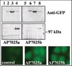
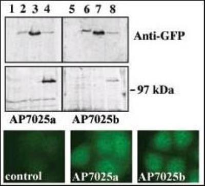
- Western Blot: PRKD3/nPKC nu Antibody [NBP2-30080] - Upper panel, western blot analysis of GFP fusion protein expression in Panc-1 cells by using an anti-GFP antibody. Lanes 1 and 5: non-transfected cells; lanes 2 and 6: GFP-PKD-transfected cells; lanes 3 and 7: GFP-PKD2-transfected cells; lanes 4 and 8: GFP-PKD3 transfected cells. Center panel, western blot analysis of GFP fusion protein expression in Panc-1 cells by using PKD3 N-term (AP7025a) and C-term (AP7025b) antibodies. Lower panel, indirect immunofluorescence analysis of GFP-PKD3 fusion protein expression in Panc-1 cells by using AP7025a and AP7025b antibodies. Data courtesy of Dr. Osvaldo Rey, University of California Los Angeles.
- Submitted by
- Novus Biologicals (provider)
- Main image
- Experimental details
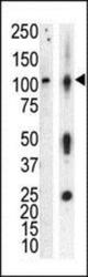
- Western Blot: PRKD3/nPKC nu Antibody [NBP2-30080] - Anti-PKCnu Pab (NBP2-30080) in lysate of HL60 cells stimulated with PMA (lane A) and mouse brain tissue lysate (lane B). PKCnu (arrow) was detected using purified Pab. Secondary HRP-anti-rabbit was used for signal visualization with chemiluminescence.

Supportive validation
- Submitted by
- Novus Biologicals (provider)
- Main image
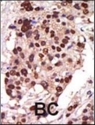
- Experimental details
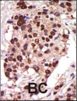
- Immunohistochemistry-Paraffin: PRKD3/nPKC nu Antibody [NBP2-30080] - Formalin-fixed and paraffin-embedded human cancer tissue reacted with the primary antibody, which was peroxidase-conjugated to the secondary antibody, followed by AEC staining. This data demonstrates the use of this antibody for immunohistochemistry; clinical relevance has not been evaluated. BC = breast carcinoma; HC = hepatocarcinoma.