Explore
 Validate
 Learn
Learn Western blot
 Immunocytochemistry
Antibody data
- Antibody Data
- Antigen structure
- References [0]
- Comments [0]
- Validations
- Western blot [2]
Submit
Validation data
Reference
Comment
Report error
- Product number
- AP13555PU-N - Provider product page

- Provider
- Acris Antibodies GmbH
- Proper citation
- Acris Antibodies GmbH Cat#AP13555PU-N, RRID:AB_1770589
- Product name
- anti PRKD3 (Center)
- Antibody type
- Polyclonal
- Antigen
- This antibody is generated from rabbits immunized with a KLH conjugated synthetic peptide selected from the center region of human PKC nu.
- Reactivity
- Human
- Host
- Rabbit
- Vial size
- 0.4 ml
- Concentration
- lot specific
No comments: Submit comment
Supportive validation
- Submitted by
- Acris Antibodies GmbH (provider)
- Main image
- Experimental details
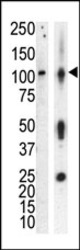
- Western blot analysis of anti-PKCnu Pab in lysate of HL60 cells stimulated with PMA (lane A) and mouse brain tissue lysate (lane B). PKCnu (arrow) was detected using purified Pab. Secondary HRP-anti-rabbit was used for signal visualization with chemiluminescence.

- Submitted by
- Acris Antibodies GmbH (provider)
- Main image

- Experimental details
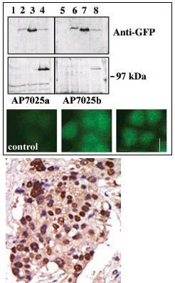
- TOP: Upper panel, western blot analysis of GFP fusion protein expression in Panc-1 cells by using an anti-GFP antibody. Lanes 1 and 5: non-transfected cells; lanes 2 and 6: GFP-PKD-transfected cells; lanes 3 and 7: GFP-PKD2-transfected cells; lanes 4 and 8: GFP-PKD3 transfected cells. Center panel, western blot analysis of GFP fusion protein expression in Panc-1 cells by using PKD3 N-term (AP13555PU-N) and C-term (AP13556PU-N) antibodies. Lower panel, indirect immunofluorescence analysis of GFP-PKD3 fusion protein expression in Panc-1 cells by using AP13555PU-N and AP13556PU-N antibodies. BOTTOM: Formalin-fixed and paraffin-embedded human cancer tissue reacted with the primary antibody, which was peroxidase-conjugated to the secondary antibody, followed by AEC staining.