Explore
 Validate
 Learn
Learn Western blot
Antibody data
- Antibody Data
- Antigen structure
- References [0]
- Comments [0]
- Validations
- Western blot [2]
- Immunocytochemistry [1]
- Immunohistochemistry [1]
Submit
Validation data
Reference
Comment
Report error
- Product number
- GTX25285 - Provider product page

- Provider
- GeneTex
- Proper citation
- GeneTex Cat#GTX25285, RRID:AB_380495
- Product name
- PKC nu antibody
- Antibody type
- Polyclonal
- Reactivity
- Human, Mouse
- Host
- Rabbit
No comments: Submit comment
Supportive validation
- Submitted by
- GeneTex (provider)
- Main image

- Experimental details
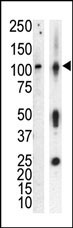
- Western blot analysis of anti-PKCnu Pab (GTX25285) in lysate of HL60 cells stimulated with PMA (lane A) and mouse brain tissue lysate (lane B). PKCnu (arrow) was detected using purified Pab. Secondary HRP-anti-rabbit was used for signal visualization with chemiluminescence.
- Submitted by
- GeneTex (provider)
- Main image
- Experimental details
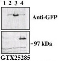
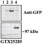
- Upper panel, western blot analysis of GFP fusion protein expression in Panc-1 cells by using an anti-GFP antibody.Lane 1: non-transfected cellsLane 2: GFP-PKD-transfected cellsLane 3: GFP-PKD2-transfected cellsLane 4: GFP-PKD3 transfected cellsCenter panel, western blot analysis of GFP fusion protein expression in Panc-1 cells by using PKD3 N-term antibody (GTX25285).
Supportive validation
- Submitted by
- GeneTex (provider)
- Main image

- Experimental details
- Indirect immunofluorescence analysis of GFP-PKD3 fusion protein expression in Panc-1 cells by using PKD3 N-term (GTX25285) antibody.

Supportive validation
- Submitted by
- GeneTex (provider)
- Main image

- Experimental details
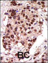
- Formalin-fixed and paraffin-embedded human cancer tissue reacted with the primary PKD3 N-term antibody (GTX25285), which was peroxidase-conjμgated to the secondary antibody, followed by AEC staining. This data demonstrates the use of this antibody for immunohistochemistry.BC = breast carcinoma