Explore
 Validate
 Learn
Learn Western blot
Antibody data
- Antibody Data
- Antigen structure
- References [3]
- Comments [0]
- Validations
- Western blot [2]
Submit
Validation data
Reference
Comment
Report error
- Product number
- AF1835 - Provider product page

- Provider
- R&D Systems
- Product name
- Mouse Semaphorin 7A Antibody
- Antibody type
- Polyclonal
- Description
- Antigen Affinity-purified. Detects mouse Semaphorin 7A in direct ELISAs and Western blots. In these formats, less than 1% cross-reactivity with recombinant mouse (rm) Semaphorin 3C, rmSemaphorin 3E, rmSemaphorin 3F, rmSemaphorin 6A, rmSemaphorin 6B, rmSemaphorin 6C, recombinant human (rh) Semaphorin 3A, rhSemaphorin 3B, and rhSemaphorin 6D is observed.
- Reactivity
- Mouse
- Host
- Goat
- Conjugate
- Unconjugated
- Antigen sequence
Q9QUR8- Isotype
- IgG
- Vial size
- 100 ug
- Concentration
- LYOPH
- Storage
- Use a manual defrost freezer and avoid repeated freeze-thaw cycles. 12 months from date of receipt, -20 to -70 °C as supplied. 1 month, 2 to 8 °C under sterile conditions after reconstitution. 6 months, -20 to -70 °C under sterile conditions after reconstitution.
Submitted references Sema7A/PlxnCl signaling triggers activity-dependent olfactory synapse formation.
Rewiring the taste system.
Semaphorin7A regulates neuroglial plasticity in the adult hypothalamic median eminence.
Inoue N, Nishizumi H, Naritsuka H, Kiyonari H, Sakano H
Nature communications 2018 May 9;9(1):1842
Nature communications 2018 May 9;9(1):1842
Rewiring the taste system.
Lee H, Macpherson LJ, Parada CA, Zuker CS, Ryba NJP
Nature 2017 Aug 17;548(7667):330-333
Nature 2017 Aug 17;548(7667):330-333
Semaphorin7A regulates neuroglial plasticity in the adult hypothalamic median eminence.
Parkash J, Messina A, Langlet F, Cimino I, Loyens A, Mazur D, Gallet S, Balland E, Malone SA, Pralong F, Cagnoni G, Schellino R, De Marchis S, Mazzone M, Pasterkamp RJ, Tamagnone L, Prevot V, Giacobini P
Nature communications 2015 Feb 27;6:6385
Nature communications 2015 Feb 27;6:6385
No comments: Submit comment
Supportive validation
- Submitted by
- R&D Systems (provider)
- Main image
- Experimental details
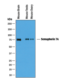
- Detection of Mouse Semaphorin 7A by Western Blot. Western blot shows lysates of mouse brain tissue, mouse testis tissue, and mouse ovary tissue. PVDF membrane was probed with 0.25 µg/mL of Goat Anti-Mouse Semaphorin 7A Antigen Affinity-purified Polyclonal Antibody (Catalog # AF1835) followed by HRP-conjugated Anti-Goat IgG Secondary Antibody (Catalog # HAF019). A specific band was detected for Semaphorin 7A at approximately 75 kDa (as indicated). This experiment was conducted under reducing conditions and using Immunoblot Buffer Group 1.
- Submitted by
- R&D Systems (provider)
- Main image

- Experimental details
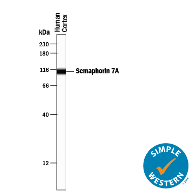
- Detection of Human Semaphorin 7A by Simple Western<SUP abp="261">TM. Simple Western lane view shows lysates of human brain (cortex) tissue, loaded at 0.2 mg/mL. A specific band was detected for Semaphorin 7A at approximately 109 kDa (as indicated) using 2.5 µg/mL of Goat Anti-Mouse Semaphorin 7A Antigen Affinity-purified Polyclonal Antibody (Catalog # AF1835) followed by 1:50 dilution of HRP-conjugated Anti-Goat IgG Secondary Antibody (Catalog # HAF109). This experiment was conducted under reducing conditions and using the 12-230 kDa separation system.