Explore
 Validate
 Learn
Learn Immunohistochemistry
Antibody data
- Antibody Data
- Antigen structure
- References [3]
- Comments [0]
- Validations
- Immunohistochemistry [1]
Submit
Validation data
Reference
Comment
Report error
- Product number
- MA5-12350 - Provider product page

- Provider
- Invitrogen Antibodies
- Product name
- MUC3 Monoclonal Antibody (1143/B7)
- Antibody type
- Monoclonal
- Antigen
- Synthetic peptide
- Description
- MA5-12350 targets Mucin 3 in IHC (P) applications and shows reactivity with Human samples.
- Antibody clone number
- 1143/B7
- Concentration
- 0.2 mg/mL
Submitted references MUC5AC, cytokeratin 20 and HER2 expression and K-RAS mutations within mucinogenic growth in congenital pulmonary airway malformations.
Development of human minor salivary glands: expression of mucins according to stage of morphogenesis.
Selective induction of mucin-3 by hypoxia in intestinal epithelia.
Rossi G, Gasser B, Sartori G, Migaldi M, Costantini M, Mengoli MC, Piccioli S, Cavazza A, Rivasi F
Histopathology 2012 Jun;60(7):1133-43
Histopathology 2012 Jun;60(7):1133-43
Development of human minor salivary glands: expression of mucins according to stage of morphogenesis.
Teshima TH, Ianez RF, Coutinho-Camillo CM, Buim ME, Soares FA, Lourenço SV
Journal of anatomy 2011 Sep;219(3):410-7
Journal of anatomy 2011 Sep;219(3):410-7
Selective induction of mucin-3 by hypoxia in intestinal epithelia.
Louis NA, Hamilton KE, Canny G, Shekels LL, Ho SB, Colgan SP
Journal of cellular biochemistry 2006 Dec 15;99(6):1616-27
Journal of cellular biochemistry 2006 Dec 15;99(6):1616-27
No comments: Submit comment
Supportive validation
- Submitted by
- Invitrogen Antibodies (provider)
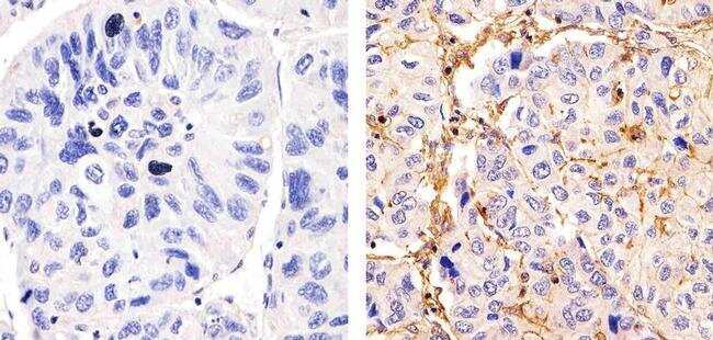
- Main image
- Experimental details
- Immunohistochemistry analysis of Mucin 3 showing staining in the membrane and weak cytoplasm of paraffin-embedded human hepatocarcinoma (right) compared to a negative control without primary antibody (left). To expose target proteins, antigen retrieval was performed using 10mM sodium citrate (pH 6.0), microwaved for 8-15 min. Following antigen retrieval, tissues were blocked in 3% H2O2-methanol for 15 min at room temperature, washed with ddH2O and PBS, and then probed with a Mucin 3 Mouse Monoclonal Antibody (Product # MA5-12350) diluted in 3% BSA-PBS at a dilution of 1:20 for 1 hour at 37°C in a humidified chamber. Tissues were washed extensively in PBST and detection was performed using an HRP-conjugated secondary antibody followed by colorimetric detection using a DAB kit. Tissues were counterstained with hematoxylin and dehydrated with ethanol and xylene to prep for mounting.