Explore
 Validate
 Learn
Learn Immunohistochemistry
Antibody data
- Antibody Data
- Antigen structure
- References [4]
- Comments [0]
- Validations
- Immunohistochemistry [12]
- Other assay [2]
Submit
Validation data
Reference
Comment
Report error
- Product number
- PA5-79702 - Provider product page

- Provider
- Invitrogen Antibodies
- Product name
- MUC2 Polyclonal Antibody
- Antibody type
- Polyclonal
- Antigen
- Synthetic peptide
- Description
- Reconstitute with 0.2 mL of distilled water to yield a concentration of 500 µg/mL. Positive Control - IHC: human colon cancer tissue, mouse colon tissue, rat colon tissue.
- Reactivity
- Human, Mouse, Rat
- Host
- Rabbit
- Isotype
- IgG
- Vial size
- 100 µg
- Concentration
- 500 µg/mL
- Storage
- -20°C
Submitted references Comparison of the host response to larvicidal and nonlarvicidal treatment of naturally acquired cyathostomin infections in horses.
Rapid establishment of human colonic organoid knockout lines.
A single cell survey of the microbial impacts on the mouse small intestinal epithelium.
Structural and functional variations in human bronchial epithelial cells cultured in air-liquid interface using different growth media.
Steuer AE, Scoggin K, Stewart JC, Barker VD, Adams AA, Loynachan AT, Nielsen MK
Parasite immunology 2022 Oct;44(10):e12941
Parasite immunology 2022 Oct;44(10):e12941
Rapid establishment of human colonic organoid knockout lines.
Gu W, Colarusso JL, Dame MK, Spence JR, Zhou Q
STAR protocols 2022 Jun 17;3(2):101308
STAR protocols 2022 Jun 17;3(2):101308
A single cell survey of the microbial impacts on the mouse small intestinal epithelium.
Tsang DKL, Wang RJ, De Sa O, Ayyaz A, Foerster EG, Bayer G, Goyal S, Trcka D, Ghoshal B, Wrana JL, Girardin SE, Philpott DJ
Gut microbes 2022 Jan-Dec;14(1):2108281
Gut microbes 2022 Jan-Dec;14(1):2108281
Structural and functional variations in human bronchial epithelial cells cultured in air-liquid interface using different growth media.
Leung C, Wadsworth SJ, Yang SJ, Dorscheid DR
American journal of physiology. Lung cellular and molecular physiology 2020 May 1;318(5):L1063-L1073
American journal of physiology. Lung cellular and molecular physiology 2020 May 1;318(5):L1063-L1073
No comments: Submit comment
Supportive validation
- Submitted by
- Invitrogen Antibodies (provider)
- Main image
- Experimental details
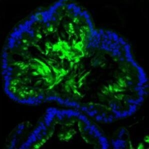
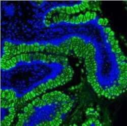
- Immunohistochemical analysis of MUC2 in paraffin-embedded section of human intestine cancer tissue. Heat mediated antigen retrieval was performed in citrate buffer (pH6, epitope retrieval solution) for 20 mins. The tissue section was blocked with 10% goat serum. The tissue section was then incubated with 5μg/mL rabbit anti-MUC2 antibody (Product # PA5-79702) overnight at 4°C. DyLight®488 Conjugated Goat Anti-Rabbit IgG was used as secondary antibody at 1:100 dilution and incubated for 30 minutes at 37°C. The section was counterstained with DAPI. Visualize using a fluorescence microscope and filter sets appropriate for the label used.

- Submitted by
- Invitrogen Antibodies (provider)
- Main image

- Experimental details
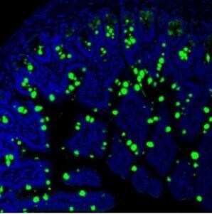
- Immunohistochemical analysis of MUC2 in paraffin-embedded section of mouse intestine tissue. Heat mediated antigen retrieval was performed in citrate buffer (pH6, epitope retrieval solution) for 20 mins. The tissue section was blocked with 10% goat serum. The tissue section was then incubated with 5μg/mL rabbit anti-MUC2 antibody (Product # PA5-79702) overnight at 4°C. DyLight®488 Conjugated Goat Anti-Rabbit IgG was used as secondary antibody at 1:100 dilution and incubated for 30 minutes at 37°C. The section was counterstained with DAPI. Visualize using a fluorescence microscope and filter sets appropriate for the label used.
- Submitted by
- Invitrogen Antibodies (provider)
- Main image
- Experimental details
- Immunohistochemical analysis of MUC2 in paraffin-embedded section of human colon organoid tissue. Heat mediated antigen retrieval was performed in citrate buffer (pH6, epitope retrieval solution) for 20 mins. The tissue section was blocked with 10% goat serum. The tissue section was then incubated with 5μg/mL rabbit anti-MUC2 antibody (Product # PA5-79702) overnight at 4°C. DyLight®488 Conjugated Goat Anti-Rabbit IgG was used as secondary antibody at 1:100 dilution and incubated for 30 minutes at 37°C. The section was counterstained with DAPI. Visualize using a fluorescence microscope and filter sets appropriate for the label used.
- Submitted by
- Invitrogen Antibodies (provider)
- Main image
- Experimental details
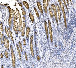
- Immunohistochemical analysis of MUC2 in a paraffin-embedded section of human colon cancer tissue. Heat mediated antigen retrieval was performed in EDTA buffer (pH 8.0, epitope retrieval solution).The tissue section was blocked with 10% goat serum. The tissue section was then incubated with 2 μg/mL rabbit anti-MUC2 antibody (Product # PA5-79702) overnight at 4°C. Biotinylated goat anti-rabbit IgG was used as secondary antibody and incubated for 30 minutes at 37°C. The tissue section was developed using Strepavidin-Biotin-Complex (SABC) with DAB as the chromogen.
- Submitted by
- Invitrogen Antibodies (provider)
- Main image
- Experimental details
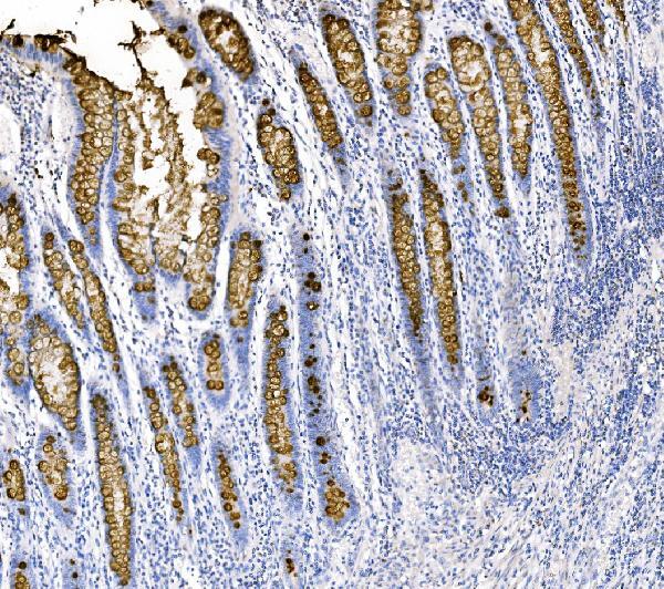
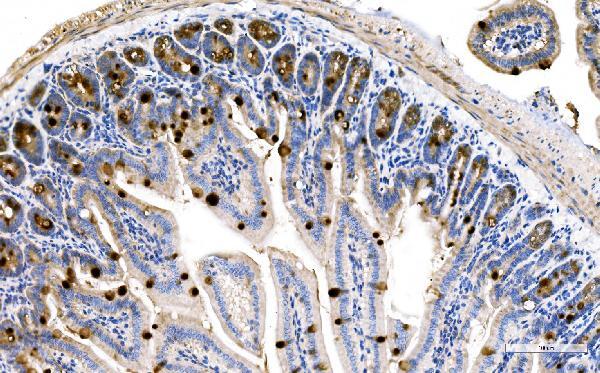
- Immunohistochemical analysis of MUC2 in a paraffin-embedded section of mouse colon tissue. Heat mediated antigen retrieval was performed in EDTA buffer (pH 8.0, epitope retrieval solution).The tissue section was blocked with 10% goat serum. The tissue section was then incubated with 2 μg/mL rabbit anti-MUC2 antibody (Product # PA5-79702) overnight at 4°C. Biotinylated goat anti-rabbit IgG was used as secondary antibody and incubated for 30 minutes at 37°C. The tissue section was developed using Strepavidin-Biotin-Complex (SABC) with DAB as the chromogen.
- Submitted by
- Invitrogen Antibodies (provider)
- Main image

- Experimental details
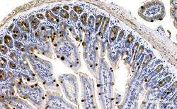
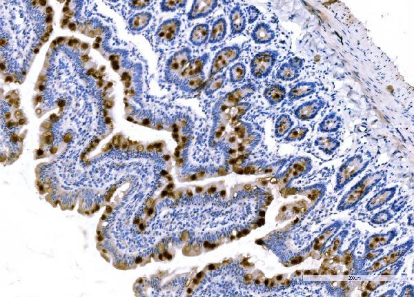
- Immunohistochemical analysis of MUC2 in a paraffin-embedded section of rat colon tissue. Heat mediated antigen retrieval was performed in EDTA buffer (pH 8.0, epitope retrieval solution).The tissue section was blocked with 10% goat serum. The tissue section was then incubated with 2 μg/mL rabbit anti-MUC2 antibody (Product # PA5-79702) overnight at 4°C. Biotinylated goat anti-rabbit IgG was used as secondary antibody and incubated for 30 minutes at 37°C. The tissue section was developed using Strepavidin-Biotin-Complex (SABC) with DAB as the chromogen.
- Submitted by
- Invitrogen Antibodies (provider)
- Main image

- Experimental details
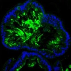
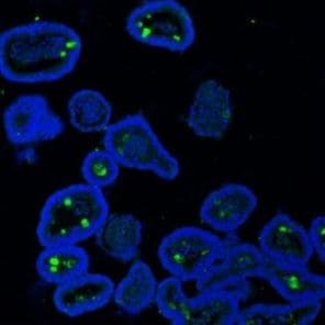
- Immunohistochemical analysis of MUC2 in paraffin-embedded section of mouse ileum tissue. Heat mediated antigen retrieval was performed in citrate buffer (pH6, epitope retrieval solution) for 20 mins. The tissue section was blocked with 10% goat serum. The tissue section was then incubated with 5μg/mL rabbit anti-MUC2 antibody (Product # PA5-79702) overnight at 4°C. DyLight®488 Conjugated Goat Anti-Rabbit IgG was used as secondary antibody at 1:100 dilution and incubated for 30 minutes at 37°C. The section was counterstained with DAPI. Visualize using a fluorescence microscope and filter sets appropriate for the label used.
- Submitted by
- Invitrogen Antibodies (provider)
- Main image
- Experimental details
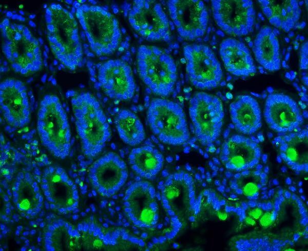
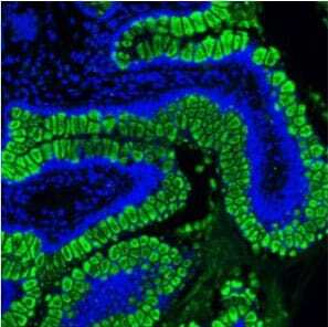
- Immunohistochemistry (Paraffin) analysis of MUC2 in paraffin-embedded section of human ileum tissue using MUC2 Polyclonal Antibody (Product # PA5-79702). Heat mediated antigen retrieval was performed in citrate buffer (pH6, epitope retrieval solution ) for 20 mins. The tissue section was blocked with 10% goat serum. The tissue section was then incubated with the primary antibody at a 5 µg/mL dilution overnight at 4°C. DyLight 488 conjugated goat anti-rabbit IgG was used as secondary antibody at 1:100 dilution and incubated for 30 minutes at 37°C. The section was counterstained with DAPI. Visualize using a fluorescence microscope and filter sets appropriate for the label used.
- Submitted by
- Invitrogen Antibodies (provider)
- Main image
- Experimental details
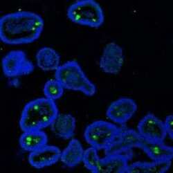
- Immunohistochemistry (Paraffin) analysis of MUC2 in paraffin-embedded section of mouse ileum organoid tissue using MUC2 Polyclonal Antibody (Product # PA5-79702). Heat mediated antigen retrieval was performed in citrate buffer (pH6, epitope retrieval solution ) for 20 mins. The tissue section was blocked with 10% goat serum. The tissue section was then incubated with the primary antibody at a 5 µg/mL dilution overnight at 4°C. DyLight 488 conjugated goat anti-rabbit IgG was used as secondary antibody at 1:100 dilution and incubated for 30 minutes at 37°C. The section was counterstained with DAPI. Visualize using a fluorescence microscope and filter sets appropriate for the label used.
- Submitted by
- Invitrogen Antibodies (provider)
- Main image
- Experimental details
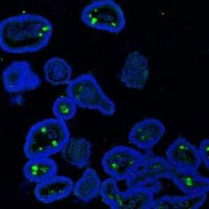
- Immunohistochemical analysis of MUC2 in paraffin-embedded section of mouse ileum organoid tissue. Heat mediated antigen retrieval was performed in citrate buffer (pH6, epitope retrieval solution) for 20 mins. The tissue section was blocked with 10% goat serum. The tissue section was then incubated with 5μg/mL rabbit anti-MUC2 antibody (Product # PA5-79702) overnight at 4°C. DyLight®488 Conjugated Goat Anti-Rabbit IgG was used as secondary antibody at 1:100 dilution and incubated for 30 minutes at 37°C. The section was counterstained with DAPI. Visualize using a fluorescence microscope and filter sets appropriate for the label used.
- Submitted by
- Invitrogen Antibodies (provider)
- Main image
- Experimental details
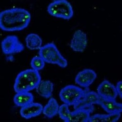
- Immunohistochemistry (Paraffin) analysis of MUC2 in paraffin-embedded section of mouse ileum tissue using MUC2 Polyclonal Antibody (Product # PA5-79702). Heat mediated antigen retrieval was performed in citrate buffer (pH6, epitope retrieval solution ) for 20 mins. The tissue section was blocked with 10% goat serum. The tissue section was then incubated with the primary antibody at a 5 µg/mL dilution overnight at 4°C. DyLight 488 conjugated goat anti-rabbit IgG was used as secondary antibody at 1:100 dilution and incubated for 30 minutes at 37°C. The section was counterstained with DAPI. Visualize using a fluorescence microscope and filter sets appropriate for the label used.
- Submitted by
- Invitrogen Antibodies (provider)
- Main image

- Experimental details
- Immunohistochemistry (Paraffin) analysis of MUC2 in paraffin-embedded section of human colon organoid tissue using MUC2 Polyclonal Antibody (Product # PA5-79702). Heat mediated antigen retrieval was performed in citrate buffer (pH6, epitope retrieval solution ) for 20 mins. The tissue section was blocked with 10% goat serum. The tissue section was then incubated with the primary antibody at a 5 µg/mL dilution overnight at 4°C. DyLight 488 conjugated goat anti-rabbit IgG was used as secondary antibody at 1:100 dilution and incubated for 30 minutes at 37°C. The section was counterstained with DAPI. Visualize using a fluorescence microscope and filter sets appropriate for the label used.

Supportive validation
- Submitted by
- Invitrogen Antibodies (provider)
- Main image

- Experimental details
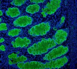
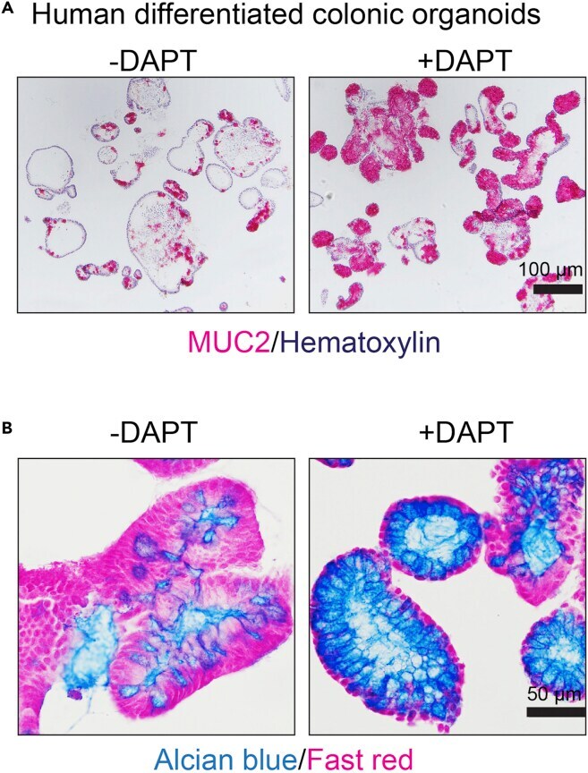
- Figure 6 DAPT promotes goblet cell differentiation (A) Representative images of differentiated organoid with or without DAPT stained with anti-MUC2 antibody (Red) and Hematoxylin nuclear stain (blue). (B) Representative images of differentiated organoids with or without DAPT stained with Alcian blue (blue) and fast red (red).
- Submitted by
- Invitrogen Antibodies (provider)
- Main image

- Experimental details
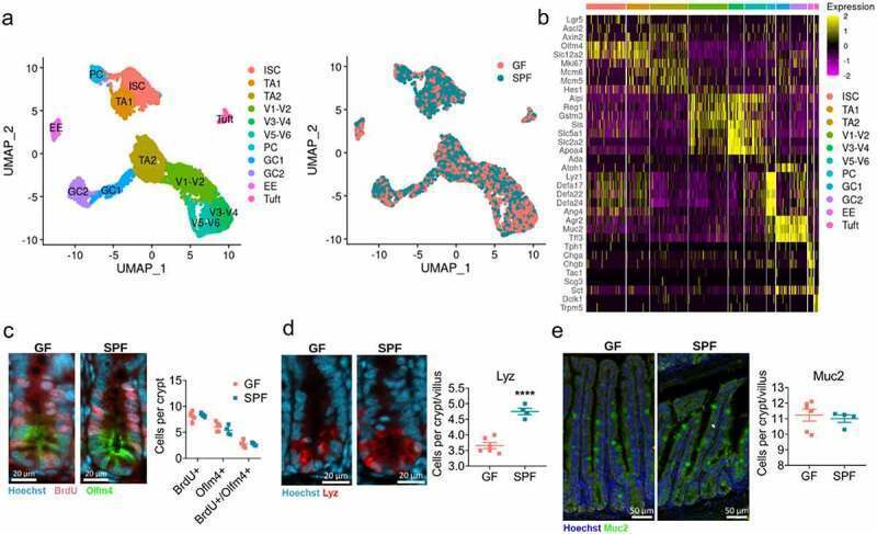
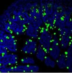
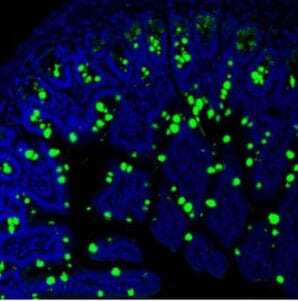
- Figure 1. Overview of the cellular landscape of the GF and SPF small intestinal epithelium . A) scRNAseq cell clusters and distribution of integrated GF and SPF epithelial cells. B) Heatmap of marker genes used to annotate epithelial cell subsets C) BrdU and Oflm4, D) Lyz, E) Muc2 staining and quantification of jejunal/ileal small intestinal crypts from GF and SPF mice. Each point represents the average counts of 20 crypts or crypt-villus axis from one mouse (n = 4-6). Representative images shown. Bars represent mean +- SEM. ****, p < .0001 by two-tailed Student's t-test.