 Explore
 Validate
 Learn
Learn Western blot
 Flow cytometry
Antibody data
- Antibody Data
- Antigen structure
- References [0]
- Comments [0]
- Validations
- Western blot [3]
- Immunoprecipitation [1]
- Immunohistochemistry [2]
Submit
Validation data
Reference
Comment
Report error
- Product number
- TA328682 - Provider product page

- Provider
- OriGene
- Product name
- Rabbit Polyclonal Anti-Human p75NTR(extracellular)
- Antibody type
- Polyclonal
- Description
- Rabbit Polyclonal Anti-Human p75NTR(extracellular)
- Host
- Rabbit
- Conjugate
- Unconjugated
- Epitope
- NGFR
- Antibody clone number
- NULL
- Vial size
- 200 µl
- Concentration
- NULL
No comments: Submit comment
Supportive validation
- Submitted by
- OriGene (provider)
- Main image
- Experimental details
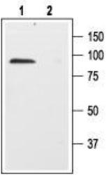
- Western blot analysis of rat brain membranes: 1. Anti-Human p75NTR (extracellular) antibody, (1:200). 2. Anti-Human p75NTR (extracellular) antibody, preincubated with the control peptide antigen.
- Validation comment
- WB
- Submitted by
- OriGene (provider)
- Main image

- Experimental details
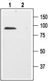
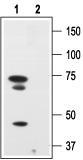
- Western blot analysis of human melanoma cells A875: 1. Anti-Human p75NTR (extracellular) antibody, (1:200). 2. Anti-Human p75NTR (extracellular) antibody, preincubated with the control peptide.
- Validation comment
- WB
- Submitted by
- OriGene (provider)
- Main image

- Experimental details
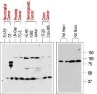
- WB analysis of normal rat tissue (right) and in human cancer cell lines (left): p75NTR is visualized with Anti-Human p75NTR (extracellular) antibody , (1:200). Interestingly, p75NTR from rat (right blot and the C6 cell line) and human (left blot) samples run with a different apparent MW, probably due to species-specific differential glycosylation.
- Validation comment
- WB
Supportive validation
- Submitted by
- OriGene (provider)
- Main image

- Experimental details
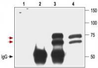
- Immunoprecipitation of 3T3/p75NTR transfected cells: 1. Cell lysate + protein A beads. 2. Cell lysate + protein A beads + pre-immune rabbit serum. 3. Cell lysate + protein A beads + Anti-Human p75NTR (extracellular) antibody. 4. Cell lysate. Red arrows indicate the p75NTR receptor while the black arrow shows the IgG heavy chain. Immunoblot was performed with the Anti-Human p75NTR (extracellular) antibody.
- Validation comment
- IP
Supportive validation
- Submitted by
- OriGene (provider)
- Main image

- Experimental details
- Expression of p75NTR in rat brain. Immunohistochemical staining of rat brain with Anti-Human p75NTR (extracellular) antibody. A. Cells in the diagonal band are stained positive for p75NTR. B. Staining of the same section with goat anti-ChAT confirms that p75NTR staining is specific to cholinergic neurons.
- Validation comment
- IHC

- Submitted by
- OriGene (provider)
- Main image

- Experimental details
- Expression of p75NTR in rat brain. Immunohistochemical staining of rat brain with Anti-Human p75NTR (extracellular) antibody. A. Cells in the nucleus basalis mangocellularis are stained positive for p75NTR. B. Staining of the same section with goat anti-ChAT confirms that the p75NTR staining is specific to cholinergic neurons.
- Validation comment
- IHC
