 Explore
 Validate
 Learn
Learn Western blot
Antibody data
- Antibody Data
- Antigen structure
- References [0]
- Comments [0]
- Validations
- Western blot [3]
- Immunocytochemistry [1]
- Immunohistochemistry [2]
Submit
Validation data
Reference
Comment
Report error
- Product number
- TA328684 - Provider product page

- Provider
- OriGene
- Product name
- Rabbit Polyclonal Anti-Neurotensin Receptor 2
- Antibody type
- Polyclonal
- Description
- Rabbit Polyclonal Anti-Neurotensin Receptor 2
- Host
- Rabbit
- Conjugate
- Unconjugated
- Epitope
- NGFR
- Antibody clone number
- NULL
- Vial size
- 200 µl
- Concentration
- NULL
No comments: Submit comment
Supportive validation
- Submitted by
- OriGene (provider)
- Main image

- Experimental details
- Western blot analysis of rat dorsal root ganglion lysates: 1. Anti-Neurotensin Receptor 2 antibody, (1:200). 2. Anti-Neurotensin Receptor 2 antibody, preincubated with the control peptide antigen.
- Validation comment
- WB

- Submitted by
- OriGene (provider)
- Main image

- Experimental details
- Western blot analysis of rat brain lysates: 1. Anti-Neurotensin Receptor 2 antibody, (1:200). 2. Anti-Neurotensin Receptor 2 antibody, preincubated with the control peptide antigen.
- Validation comment
- WB

- Submitted by
- OriGene (provider)
- Main image

- Experimental details
- Western blot analysis of mouse brain lysates: 1. Anti-Neurotensin Receptor 2 antibody, (1:400). 2. Anti-Neurotensin Receptor 2 antibody, preincubated with the control peptide antigen.
- Validation comment
- WB

Supportive validation
- Submitted by
- OriGene (provider)
- Main image

- Experimental details
- Expression of NTR2 in rat C6 glioma cells. Immunocytochemical staining of rat C6 glioma cells. A. Paraformaldehyde fixed and permeabilized rat C6 glioma cells were stained with Anti-Neurotensin Receptor 2 antibody, (1:200), followed by Alexa-555 conjugated goat-anti-rabbit- secondary antibody (orange staining). Nuclei were visualized with the cell permeable dye Hoechst 33342 (blue staining). B. Live view of the same field as in (A).
- Validation comment
- IF

Supportive validation
- Submitted by
- OriGene (provider)
- Main image

- Experimental details
- IHC staining of perfusion-fixed brain frozen rat mediobasal (MB) hypothalamus sections using Anti-Neurotensin Receptor 2 antibody, (1:100), (green). A. Neurotensin Receptor 2 (NTS2) appears in neuronal fibers marked by white arrows. B. In the same section, there is staining for glial fibrillary acidic protein (red fluorescence), a marker of astrocytes. C. Merging the two images demonstrates that NTS2 is not localized in astrocytes. DAPI is used as the counterstain (blue).
- Validation comment
- IHC

- Submitted by
- OriGene (provider)
- Main image
- Experimental details
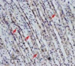
- Expression of NTR2 in rat stomach. Immunohistochemical staining of paraffin-embedded rat stomach sections using Anti-Neurotensin Receptor 2 antibody , (1:50), (brown). Staining is specific for parietal cells (arrows) of the gastric glands. Hematoxilin is used as the counterstain.
- Validation comment
- IHC
