 Explore
 Validate
 Learn
Learn Western blot
 Immunohistochemistry
Antibody data
- Antibody Data
- Antigen structure
- References [7]
- Comments [0]
- Validations
- Immunohistochemistry [1]
- Other assay [3]
Submit
Validation data
Reference
Comment
Report error
- Product number
- 14-9400-80 - Provider product page

- Provider
- Invitrogen Antibodies
- Product name
- CD271 (NGF Receptor) Monoclonal Antibody (ME20.4), eBioscience™
- Antibody type
- Monoclonal
- Antigen
- Other
- Description
- Description: The monoclonal antibody ME20.4 recognizes CD271, a 75 kDa type I transmembrane protein that belongs to the low-affinity neurotrophin receptor and tumor necrosis factor receptor superfamily. CD271 is expressed by neurons, Schwann cells, melanocytes, mesenchymal stem/stromal cells, neural crest stem cells, astrocytes, oligodendrocytes, and a subset of lymphoid cells including follicular dendritic cells. CD271 binds to the ligands NGF, BDNF, NTF3, and NTF4. CD271 contains a death domain which induces apoptosis and regulates cell growth and proliferation through interactions with TRAF2, TRAF4, TRAF6, PTPN13, and RANBP9. The ME20.4 monoclonal antibody recognizes human, non-human primate, mouse, rat, sheep, rabbit, cat, dog, and pig CD271. Applications Reported: This ME20.4 antibody has been reported for use in flow cytometric analysis, western blotting, microscopy, immunohistochemical staining, and immunocytochemistry. Applications Tested: This ME20.4 antibody has been tested by flow cytometric analysis of SK-N-SH cells. This can be used at less than or equal to 0.25 µg per test. A test is defined as the amount (µg) of antibody that will stain a cell sample in a final volume of 100 µL. Cell number should be determined empirically but can range from 10^5 to 10^8 cells/test. This ME20.4 antibody has also been tested by immunohistochemistry of formalin-fixed paraffin embedded human tissue using high pH antigen retrieval and can be used at less than or equal to 5 µg/mL. It is recommended that the antibody be carefully titrated for optimal performance in the assay of interest. Purity: Greater than 90%, as determined by SDS-PAGE. Aggregation: Less than 10%, as determined by HPLC. Filtration: 0.2 µm post-manufacturing filtered.
- Reactivity
- Human, Mouse, Rat, Canine, Porcine, Rabbit
- Host
- Mouse
- Isotype
- IgG
- Antibody clone number
- ME20.4
- Vial size
- 25 µg
- Concentration
- 0.5 mg/mL
- Storage
- 4° C
Submitted references Widespread airway distribution and short-term phenotypic correction of cystic fibrosis pigs following aerosol delivery of piggyBac/adenovirus.
IL-1β-induces NF-κB and upregulates microRNA-372 to inhibit spinal cord injury recovery.
Ibuprofen inhibits survival of bladder cancer cells by induced expression of the p75NTR tumor suppressor protein.
Nerve growth factor induces the re-expression of functional androgen receptors and p75(NGFR) in the androgen-insensitive prostate cancer cell line DU145.
Bone marrow stroma in humans: anti-nerve growth factor receptor antibodies selectively stain reticular cells in vivo and in vitro.
Expression of human nerve growth factor receptor on cells derived from all three germ layers.
Characterization of nerve growth factor receptor in neural crest tumors using monoclonal antibodies.
Cooney AL, Singh BK, Loza LM, Thornell IM, Hippee CE, Powers LS, Ostedgaard LS, Meyerholz DK, Wohlford-Lenane C, Stoltz DA, B McCray P Jr, Sinn PL
Nucleic acids research 2018 Oct 12;46(18):9591-9600
Nucleic acids research 2018 Oct 12;46(18):9591-9600
IL-1β-induces NF-κB and upregulates microRNA-372 to inhibit spinal cord injury recovery.
Zhou W, Yuan T, Gao Y, Yin P, Liu W, Pan C, Liu Y, Yu X
Journal of neurophysiology 2017 Jun 1;117(6):2282-2291
Journal of neurophysiology 2017 Jun 1;117(6):2282-2291
Ibuprofen inhibits survival of bladder cancer cells by induced expression of the p75NTR tumor suppressor protein.
Khwaja F, Allen J, Lynch J, Andrews P, Djakiew D
Cancer research 2004 Sep 1;64(17):6207-13
Cancer research 2004 Sep 1;64(17):6207-13
Nerve growth factor induces the re-expression of functional androgen receptors and p75(NGFR) in the androgen-insensitive prostate cancer cell line DU145.
Sigala S, Tognazzi N, Rizzetti MC, Faraoni I, Missale C, Bonmassar E, Spano P
European journal of endocrinology 2002 Sep;147(3):407-15
European journal of endocrinology 2002 Sep;147(3):407-15
Bone marrow stroma in humans: anti-nerve growth factor receptor antibodies selectively stain reticular cells in vivo and in vitro.
Cattoretti G, Schiró R, Orazi A, Soligo D, Colombo MP
Blood 1993 Apr 1;81(7):1726-38
Blood 1993 Apr 1;81(7):1726-38
Expression of human nerve growth factor receptor on cells derived from all three germ layers.
Thomson TM, Rettig WJ, Chesa PG, Green SH, Mena AC, Old LJ
Experimental cell research 1988 Feb;174(2):533-9
Experimental cell research 1988 Feb;174(2):533-9
Characterization of nerve growth factor receptor in neural crest tumors using monoclonal antibodies.
Ross AH, Grob P, Bothwell M, Elder DE, Ernst CS, Marano N, Ghrist BF, Slemp CC, Herlyn M, Atkinson B
Proceedings of the National Academy of Sciences of the United States of America 1984 Nov;81(21):6681-5
Proceedings of the National Academy of Sciences of the United States of America 1984 Nov;81(21):6681-5
No comments: Submit comment
Supportive validation
- Submitted by
- Invitrogen Antibodies (provider)
- Main image

- Experimental details
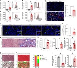
- Immunohistochemistry of formalin-fixed paraffin embedded human prostate tissue stained with 5 µg/mL of Mouse IgG1 K Isotype Control Purified (left) or 5 µg/mL of Anti-CD271 (NGF Receptor) Purified (right) followed by Anti-Mouse IgG Biotin, Streptavidin HRP, and DAB visualization.Nuclei are counterstained with hematoxylin.

Supportive validation
- Submitted by
- Invitrogen Antibodies (provider)
- Main image

- Experimental details
- Figure 4. piggyBac /Ad-GFP transduces multiple cell types. Tissue from each region of the pig lung (as depicted in Figure 1B ) was sectioned, counterstained with DAPI, and imaged. ( A and B ) piggyBac /Ad-GFP transduces submucosal glands (SMGs) throughout the trachea. ( B and C ) Tissues stained with acetyl-tubulin (red) show that piggyBac /Ad-GFP transduces ciliated (c) and non-ciliated (nc) cells at the airway surface. ( D - F ) piggyBac /Ad-GFP co-localizes with CK5 positive basal cells (red) within submucosal glands ( n = 6). Scale bars for A = 250 mum; B-F = 500 mum. ( G and H ) piggyBac /Ad-GFP transduces basal cells at the airway surface. Arrows indicate GFP positive cells that co-localize with red (G) NGFR or (H) CK14 immunostaining. Images acquired at 63x magnification ( n = 6).

- Submitted by
- Invitrogen Antibodies (provider)
- Main image
- Experimental details
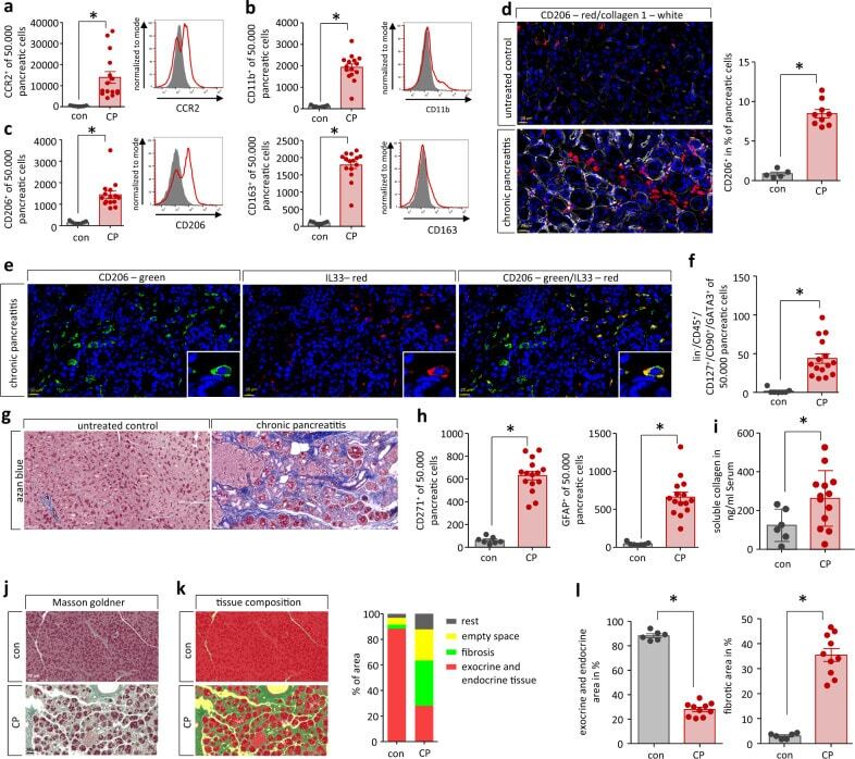
- Chronic pancreatitis leads to pancreatic tissue remodeling. a Flow cytometry analysis of isolated leukocytes from pancreatic tissue showed a significant increase of CCR2 + cells ( p = 0.0032, con n = 7/CP n = 15). b CD11b + labeling identified that also cells of the innate immune system infiltrate into the damaged pancreas ( p < 0.0001, con n = 7/CP n = 15). c Most of these cells were positive for CD206 ( p < 0.0001, con n = 7/CP n = 15) or CD163 ( p < 0.0001, con n = 7/CP n = 15), which characterized them as alternatively activated macrophages. d Labeling of CP tissue for CD206 illustrates their increase in pancreatic tissue of mice. A quantative analysis showed that ~9% of all cells were positive for CD206 in CP tissue ( p < 0.0001, con n = 5/CP n = 9), scale bars represent 20 um. e IL-33 expression of CD206 + macrophages was demonstrated by immunofluorescent labeling, scale bars represent 20 um. f This cytokine is a strong inducer of ILC2s (lin - CD45 + CD127 + CD90 + GATA3 + ) which were also significantly elevated in the pancreas of CP mice ( p = 0.0002, con n = 7/CP n = 15). g The replacement of exocrine pancreas by fibrotic tissue in CP was illustrated by azan blue staining of the pancreas, scale bars represent 50 um. h Flow cytometry analysis of isolated pancreatic cells for PSC marker proteins CD271 ( p < 0.0001, con n = 7/CP n = 15) and GFAP ( p < 0.0001, con n = 7/CP n = 15) identified a significant increase. i Serum collagen levels were significantly elevated in C
- Submitted by
- Invitrogen Antibodies (provider)
- Main image

- Experimental details
- Evaluation of the CD marker location in the synovial membrane. Immunohistochemical analysis using DAB (3, 3'-diaminobenzidine, orange/brown staining) was performed to identify the location of CD90, CD44, CD73, CD271, and CD34 expression in the normal (circle) and osteoarthritic (OA, square) synovial membrane. Representative images, containing both fibrous and adipose type synovial membrane, were chosen. The scale bar is set at 50 um. Quantification of the DAB staining was performed with Image ProPlus 6.0 software (Media Cybernetics) in 3 to 6 random regions of interest (ROI) per donor, resulting in a mean percentage of DAB positive staining areas for each donor
