 Explore
 Validate
 Learn
Learn Western blot
 Immunocytochemistry
Antibody data
- Antibody Data
- Antigen structure
- References [22]
- Comments [0]
- Validations
- Immunocytochemistry [6]
- Flow cytometry [5]
- Other assay [4]
Submit
Validation data
Reference
Comment
Report error
- Product number
- MA5-13314 - Provider product page

- Provider
- Invitrogen Antibodies
- Product name
- NGFR Monoclonal Antibody (NGFR5)
- Antibody type
- Monoclonal
- Antigen
- Other
- Description
- MA5-13314 targets Neurotrophin Receptor /NGF-Receptor (p75NGFR) in FACS, ICC/IF and WB applications and shows reactivity with Feline, Ferret, Human, Rat, Non-human primate, and Rabbit samples. This antibody does not react with rat tissue in Western blot applications. The MA5-13314 immunogen is nGFR from A875 melanoma cells.
- Reactivity
- Human, Rat, Feline, Rabbit
- Host
- Mouse
- Isotype
- IgG
- Antibody clone number
- NGFR5
- Vial size
- 500 μL
- Concentration
- 0.2 mg/mL
- Storage
- 4°C
Submitted references A genetic compensatory mechanism regulated by Jun and Mef2d modulates the expression of distinct class IIa Hdacs to ensure peripheral nerve myelination and repair.
Derivation of Airway Basal Stem Cells from Human Pluripotent Stem Cells.
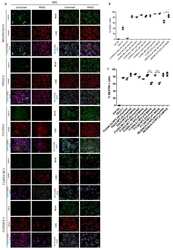
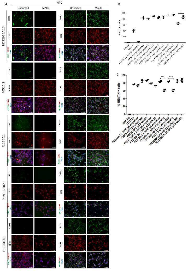
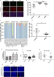
Reduced variability of neural progenitor cells and improved purity of neuronal cultures using magnetic activated cell sorting.
In vitro effect of nerve growth factor on the main traits of rabbit sperm.
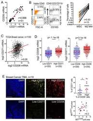
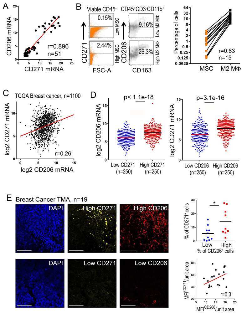
Exosomes Produced by Mesenchymal Stem Cells Drive Differentiation of Myeloid Cells into Immunosuppressive M2-Polarized Macrophages in Breast Cancer.
A phase1 study of stereotactic gene delivery of AAV2-NGF for Alzheimer's disease.
The dimensions and characteristics of the subepidermal nerve plexus in human skin--terminal Schwann cells constitute a substantial cell population within the superficial dermis.
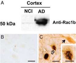
Rac1b increases with progressive tau pathology within cholinergic nucleus basalis neurons in Alzheimer's disease.
Human neurotrophin receptor p75NTR defines differentiation-oriented skeletal muscle precursor cells: implications for muscle regeneration.
Low-affinity nerve growth factor receptor (P75 NGFR) as a marker of perineural invasion in malignant melanomas.
In situ evidence of involvement of Schwann cells in ulcerative colitis: autocrine and paracrine signaling by A disintegrin and metalloprotease-17-mediated tumor necrosis factor alpha production.
Immunophenotypical pleomorphism expression in sudden cardiac death.
Immunohistochemical expression patterns of neural and neuroendocrine markers, the neural growth factor receptors and the beta-tubulin II and IV isotypes in human thymus.
The activated nerve growth factor receptor p-TrkA is selectively expressed in advanced-stage ovarian carcinoma.
Immunohistochemical localization of epithelial rests of Malassez in human periodontal membrane.
The p75 neurotrophin receptor is widely expressed in conventional papillary thyroid carcinoma.
Differential expression of the neurotrophin receptors p75NTR, TrkA, TrkB and TrkC in human peripheral blood mononuclear cells.
Origin and ontogeny of mammalian ovarian neurons.
Modulation of Fas-induced apoptosis by p75 neurotrophin receptor in a human neuroblastoma cell line.
A histological and anatomical profile of pacinian corpuscles from Dupuytren's contracture and the expression of nerve growth factor receptor.
A histological and anatomical profile of pacinian corpuscles from Dupuytren's contracture and the expression of nerve growth factor receptor.
The neurotrophins nerve growth factor, brain-derived neurotrophic factor, neurotrophin-3, and neurotrophin-4 are survival and activation factors for eosinophils in patients with allergic bronchial asthma.
Velasco-Aviles S, Patel N, Casillas-Bajo A, Frutos-Rincón L, Velasco E, Gallar J, Arthur-Farraj P, Gomez-Sanchez JA, Cabedo H
eLife 2022 Jan 25;11
eLife 2022 Jan 25;11
Derivation of Airway Basal Stem Cells from Human Pluripotent Stem Cells.
Hawkins FJ, Suzuki S, Beermann ML, Barillà C, Wang R, Villacorta-Martin C, Berical A, Jean JC, Le Suer J, Matte T, Simone-Roach C, Tang Y, Schlaeger TM, Crane AM, Matthias N, Huang SXL, Randell SH, Wu J, Spence JR, Carraro G, Stripp BR, Rab A, Sorsher EJ, Horani A, Brody SL, Davis BR, Kotton DN
Cell stem cell 2021 Jan 7;28(1):79-95.e8
Cell stem cell 2021 Jan 7;28(1):79-95.e8
Reduced variability of neural progenitor cells and improved purity of neuronal cultures using magnetic activated cell sorting.
Bowles KR, Tcw J, Qian L, Jadow BM, Goate AM
PloS one 2019;14(3):e0213374
PloS one 2019;14(3):e0213374
In vitro effect of nerve growth factor on the main traits of rabbit sperm.
Castellini C, Mattioli S, Dal Bosco A, Collodel G, Pistilli A, Stabile AM, Macchioni L, Mancuso F, Luca G, Rende M
Reproductive biology and endocrinology : RB&E 2019 Nov 12;17(1):93
Reproductive biology and endocrinology : RB&E 2019 Nov 12;17(1):93
Exosomes Produced by Mesenchymal Stem Cells Drive Differentiation of Myeloid Cells into Immunosuppressive M2-Polarized Macrophages in Breast Cancer.
Biswas S, Mandal G, Roy Chowdhury S, Purohit S, Payne KK, Anadon C, Gupta A, Swanson P, Yu X, Conejo-Garcia JR, Bhattacharyya A
Journal of immunology (Baltimore, Md. : 1950) 2019 Dec 15;203(12):3447-3460
Journal of immunology (Baltimore, Md. : 1950) 2019 Dec 15;203(12):3447-3460
A phase1 study of stereotactic gene delivery of AAV2-NGF for Alzheimer's disease.
Rafii MS, Baumann TL, Bakay RA, Ostrove JM, Siffert J, Fleisher AS, Herzog CD, Barba D, Pay M, Salmon DP, Chu Y, Kordower JH, Bishop K, Keator D, Potkin S, Bartus RT
Alzheimer's & dementia : the journal of the Alzheimer's Association 2014 Sep;10(5):571-81
Alzheimer's & dementia : the journal of the Alzheimer's Association 2014 Sep;10(5):571-81
The dimensions and characteristics of the subepidermal nerve plexus in human skin--terminal Schwann cells constitute a substantial cell population within the superficial dermis.
Reinisch CM, Tschachler E
Journal of dermatological science 2012 Mar;65(3):162-9
Journal of dermatological science 2012 Mar;65(3):162-9
Rac1b increases with progressive tau pathology within cholinergic nucleus basalis neurons in Alzheimer's disease.
Perez SE, Getova DP, He B, Counts SE, Geula C, Desire L, Coutadeur S, Peillon H, Ginsberg SD, Mufson EJ
The American journal of pathology 2012 Feb;180(2):526-40
The American journal of pathology 2012 Feb;180(2):526-40
Human neurotrophin receptor p75NTR defines differentiation-oriented skeletal muscle precursor cells: implications for muscle regeneration.
Colombo E, Romaggi S, Medico E, Menon R, Mora M, Falcone C, Lochmüller H, Confalonieri P, Mantegazza R, Morandi L, Farina C
Journal of neuropathology and experimental neurology 2011 Feb;70(2):133-42
Journal of neuropathology and experimental neurology 2011 Feb;70(2):133-42
Low-affinity nerve growth factor receptor (P75 NGFR) as a marker of perineural invasion in malignant melanomas.
Chan MM, Tahan SR
Journal of cutaneous pathology 2010 Mar;37(3):336-43
Journal of cutaneous pathology 2010 Mar;37(3):336-43
In situ evidence of involvement of Schwann cells in ulcerative colitis: autocrine and paracrine signaling by A disintegrin and metalloprotease-17-mediated tumor necrosis factor alpha production.
Mosnier JF, Jarry A, Camdessanché JP, Antoine JC, Laboisse CL
Human pathology 2009 Aug;40(8):1159-67
Human pathology 2009 Aug;40(8):1159-67
Immunophenotypical pleomorphism expression in sudden cardiac death.
Ceauşu M, Curcă C, Ardeleanu C, Dermengiu D
Romanian journal of morphology and embryology = Revue roumaine de morphologie et embryologie 2008;49(3):315-20
Romanian journal of morphology and embryology = Revue roumaine de morphologie et embryologie 2008;49(3):315-20
Immunohistochemical expression patterns of neural and neuroendocrine markers, the neural growth factor receptors and the beta-tubulin II and IV isotypes in human thymus.
Bai M, Papoudou-Bai A, Karatzias G, Doukas M, Goussia A, Stefanaki K, Rontogianni D, Dalavanga Y, Agnantis NJ, Kanavaros P
Anticancer research 2008 Jan-Feb;28(1A):295-303
Anticancer research 2008 Jan-Feb;28(1A):295-303
The activated nerve growth factor receptor p-TrkA is selectively expressed in advanced-stage ovarian carcinoma.
Ødegaard E, Staff AC, Abeler VM, Kopolovic J, Onsrud M, Lazarovici P, Davidson B
Human pathology 2007 Jan;38(1):140-6
Human pathology 2007 Jan;38(1):140-6
Immunohistochemical localization of epithelial rests of Malassez in human periodontal membrane.
Becktor KB, Nolting D, Becktor JP, Kjaer I
European journal of orthodontics 2007 Aug;29(4):350-3
European journal of orthodontics 2007 Aug;29(4):350-3
The p75 neurotrophin receptor is widely expressed in conventional papillary thyroid carcinoma.
Rocha AS, Risberg B, Magalhães J, Trovisco V, de Castro IV, Lazarovici P, Soares P, Davidson B, Sobrinho-Simões M
Human pathology 2006 May;37(5):562-8
Human pathology 2006 May;37(5):562-8
Differential expression of the neurotrophin receptors p75NTR, TrkA, TrkB and TrkC in human peripheral blood mononuclear cells.
Nassenstein C, Möhring UH, Luttmann W, Virchow JC Jr, Braun A
Experimental and toxicologic pathology : official journal of the Gesellschaft fur Toxikologische Pathologie 2006 Jun;57 Suppl 2:55-63
Experimental and toxicologic pathology : official journal of the Gesellschaft fur Toxikologische Pathologie 2006 Jun;57 Suppl 2:55-63
Origin and ontogeny of mammalian ovarian neurons.
Dees WL, Hiney JK, McArthur NH, Johnson GA, Dissen GA, Ojeda SR
Endocrinology 2006 Aug;147(8):3789-96
Endocrinology 2006 Aug;147(8):3789-96
Modulation of Fas-induced apoptosis by p75 neurotrophin receptor in a human neuroblastoma cell line.
Giraud S, Lautrette C, Bessette B, Decourt C, Mathonnet M, Jauberteau MO
Apoptosis : an international journal on programmed cell death 2005 Dec;10(6):1271-83
Apoptosis : an international journal on programmed cell death 2005 Dec;10(6):1271-83
A histological and anatomical profile of pacinian corpuscles from Dupuytren's contracture and the expression of nerve growth factor receptor.
Ehrmantant WR, Graham WP 3rd, Towfighi J, Mackay DR, Ehrlich HP
Plastic and reconstructive surgery 2004 Sep 1;114(3):721-7
Plastic and reconstructive surgery 2004 Sep 1;114(3):721-7
The neurotrophins nerve growth factor, brain-derived neurotrophic factor, neurotrophin-3, and neurotrophin-4 are survival and activation factors for eosinophils in patients with allergic bronchial asthma.
Nassenstein C, Braun A, Erpenbeck VJ, Lommatzsch M, Schmidt S, Krug N, Luttmann W, Renz H, Virchow JC Jr
The Journal of experimental medicine 2003 Aug 4;198(3):455-67
The Journal of experimental medicine 2003 Aug 4;198(3):455-67
No comments: Submit comment
Supportive validation
- Submitted by
- Invitrogen Antibodies (provider)
- Main image

- Experimental details
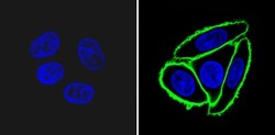
- Immunofluorescent analysis of Neurotrophin Receptor /NGF-Receptor (p75NGFR) (green) showing staining in the membrane of SW480 cells (right) compared to a negative control without primary antibody (left). Formalin-fixed cells were permeabilized with 0.1% Triton X-100 in TBS for 5-10 minutes and blocked with 3% BSA-PBS for 30 minutes at room temperature. Cells were probed with a Neurotrophin Receptor /NGF-Receptor (p75NGFR) monoclonal antibody (Product # MA5-13314) in 3% BSA-PBS at a dilution of 1:100 and incubated overnight at 4 ºC in a humidified chamber. Cells were washed with PBST and incubated with a DyLight-conjugated secondary antibody in PBS at room temperature in the dark. F-actin (red) was stained with a fluorescent red phalloidin and nuclei (blue) were stained with Hoechst or DAPI. Images were taken at a magnification of 60x.

- Submitted by
- Invitrogen Antibodies (provider)
- Main image

- Experimental details
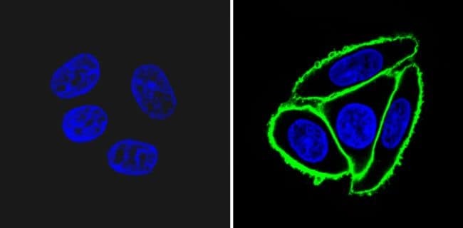
- Immunofluorescent analysis of Neurotrophin Receptor /NGF-Receptor (p75NGFR) (green) showing staining in the membrane of SW480 cells (right) compared to a negative control without primary antibody (left). Formalin-fixed cells were permeabilized with 0.1% Triton X-100 in TBS for 5-10 minutes and blocked with 3% BSA-PBS for 30 minutes at room temperature. Cells were probed with a Neurotrophin Receptor /NGF-Receptor (p75NGFR) monoclonal antibody (Product # MA5-13314) in 3% BSA-PBS at a dilution of 1:100 and incubated overnight at 4 ºC in a humidified chamber. Cells were washed with PBST and incubated with a DyLight-conjugated secondary antibody in PBS at room temperature in the dark. F-actin (red) was stained with a fluorescent red phalloidin and nuclei (blue) were stained with Hoechst or DAPI. Images were taken at a magnification of 60x.

- Submitted by
- Invitrogen Antibodies (provider)
- Main image
- Experimental details
- Immunofluorescent analysis of Neurotrophin Receptor /NGF-Receptor (p75NGFR) (green) showing staining in the membrane of SW480 cells (right) compared to a negative control without primary antibody (left). Formalin-fixed cells were permeabilized with 0.1% Triton X-100 in TBS for 5-10 minutes and blocked with 3% BSA-PBS for 30 minutes at room temperature. Cells were probed with a Neurotrophin Receptor /NGF-Receptor (p75NGFR) monoclonal antibody (Product # MA5-13314) in 3% BSA-PBS at a dilution of 1:100 and incubated overnight at 4 ºC in a humidified chamber. Cells were washed with PBST and incubated with a DyLight-conjugated secondary antibody in PBS at room temperature in the dark. F-actin (red) was stained with a fluorescent red phalloidin and nuclei (blue) were stained with Hoechst or DAPI. Images were taken at a magnification of 60x.
- Submitted by
- Invitrogen Antibodies (provider)
- Main image
- Experimental details
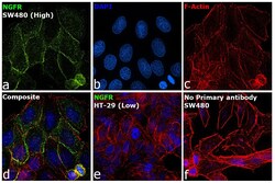
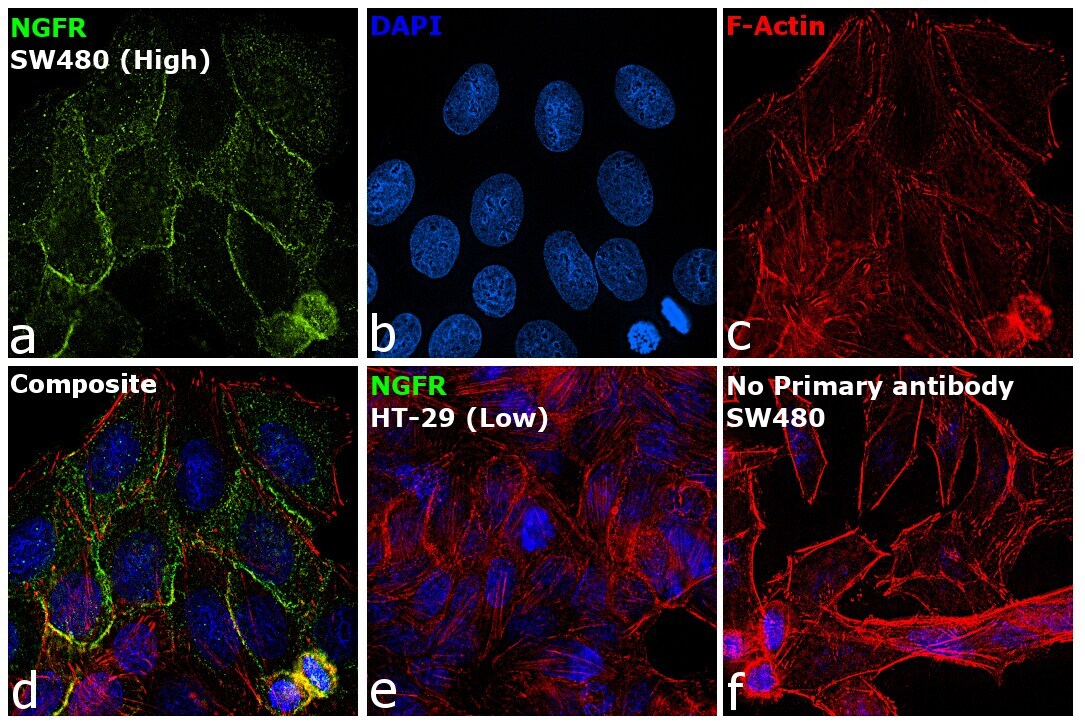
- Immunofluorescence analysis of NGFR was performed using 70% confluent log phase SW480 cells. The cells were fixed with 4% paraformaldehyde for 10 minutes, permeabilized with 0.1% Triton™ X-100 for 15 minutes, and blocked with 2% BSA for 45 minutes at room temperature. The cells were labeled with NGFR Monoclonal Antibody (NGFR5) (Product # MA5-13314) at 1:100 in 0.1% BSA, incubated at 4 degree celsius overnight and then labeled with Donkey anti-Mouse IgG (H+L) Highly Cross-Adsorbed Secondary Antibody, Alexa Fluor Plus 488 (Product # A32766), (1:2000 dilution), for 45 minutes at room temperature (Panel a: Green). Nuclei (Panel b: Blue) were stained with ProLong™ Diamond Antifade Mountant with DAPI (Product # P36962). F-actin (Panel c: Red) was stained with Rhodamine Phalloidin (Product # R415, 1:300 dilution). Panel d represents the merged image showing membranous and cytosolic localization. Panel e represents HT-29 cells showing no expression of NGFR. Panel f represents control cells with no primary antibody to assess background. The images were captured at 60X magnification.
- Submitted by
- Invitrogen Antibodies (provider)
- Main image

- Experimental details
- Immunofluorescent analysis of Neurotrophin Receptor /NGF-Receptor (p75NGFR) (green) showing staining in the membrane of SW480 cells (right) compared to a negative control without primary antibody (left). Formalin-fixed cells were permeabilized with 0.1% Triton X-100 in TBS for 5-10 minutes and blocked with 3% BSA-PBS for 30 minutes at room temperature. Cells were probed with a Neurotrophin Receptor /NGF-Receptor (p75NGFR) monoclonal antibody (Product # MA5-13314) in 3% BSA-PBS at a dilution of 1:100 and incubated overnight at 4 ºC in a humidified chamber. Cells were washed with PBST and incubated with a DyLight-conjugated secondary antibody in PBS at room temperature in the dark. F-actin (red) was stained with a fluorescent red phalloidin and nuclei (blue) were stained with Hoechst or DAPI. Images were taken at a magnification of 60x.

- Submitted by
- Invitrogen Antibodies (provider)
- Main image

- Experimental details
- Immunofluorescence analysis of NGFR was performed using 70% confluent log phase SW480 cells. The cells were fixed with 4% paraformaldehyde for 10 minutes, permeabilized with 0.1% Triton™ X-100 for 15 minutes, and blocked with 2% BSA for 45 minutes at room temperature. The cells were labeled with NGFR Monoclonal Antibody (NGFR5) (Product # MA5-13314) at 1:100 in 0.1% BSA, incubated at 4 degree celsius overnight and then labeled with Donkey anti-Mouse IgG (H+L) Highly Cross-Adsorbed Secondary Antibody, Alexa Fluor Plus 488 (Product # A32766), (1:2000 dilution), for 45 minutes at room temperature (Panel a: Green). Nuclei (Panel b: Blue) were stained with ProLong™ Diamond Antifade Mountant with DAPI (Product # P36962). F-actin (Panel c: Red) was stained with Rhodamine Phalloidin (Product # R415, 1:300 dilution). Panel d represents the merged image showing membranous and cytosolic localization. Panel e represents HT-29 cells showing no expression of NGFR. Panel f represents control cells with no primary antibody to assess background. The images were captured at 60X magnification.

Supportive validation
- Submitted by
- Invitrogen Antibodies (provider)
- Main image

- Experimental details
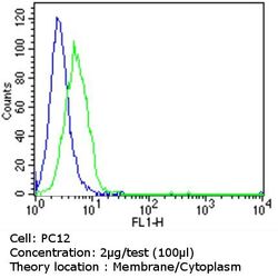
- Flow cytometry analysis of Neurotrophin Receptor /NGF-Receptor (p75NGFR) in PC12 cells (green) compared to an isotype control (blue). Cells were harvested, adjusted to a concentration of 1-5x10^6 cells/mL, fixed with 2% paraformaldehyde and washed with PBS. Cells were blocked with a 2% solution of BSA-PBS for 30 min at room temperature and incubated with a Neurotrophin Receptor /NGF-Receptor (p75NGFR) monoclonal antibody (Product # MA5-13314) at a dilution of 2 µg/test for 60 min at room temperature. Cells were then incubated for 40 min at room temperature in the dark using a Dylight 488-conjugated goat anti-mouse IgG (H+L) secondary antibody and re-suspended in PBS for FACS analysis.

- Submitted by
- Invitrogen Antibodies (provider)
- Main image
- Experimental details
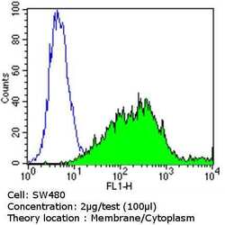
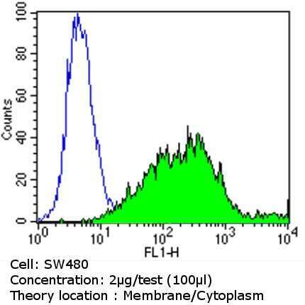
- Flow cytometry analysis of Neurotrophin Receptor /NGF-Receptor (p75NGFR) in SW480 cells (green) compared to an isotype control (blue). Cells were harvested, adjusted to a concentration of 1-5x10^6 cells/mL, fixed with 2% paraformaldehyde and washed with PBS. Cells were blocked with a 2% solution of BSA-PBS for 30 min at room temperature and incubated with a Neurotrophin Receptor /NGF-Receptor (p75NGFR) monoclonal antibody (Product # MA5-13314) at a dilution of 2 µg/test for 60 min at room temperature. Cells were then incubated for 40 min at room temperature in the dark using a Dylight 488-conjugated goat anti-mouse IgG (H+L) secondary antibody and re-suspended in PBS for FACS analysis.
- Submitted by
- Invitrogen Antibodies (provider)
- Main image
- Experimental details
- Flow cytometry analysis of Neurotrophin Receptor /NGF-Receptor (p75NGFR) in PC12 cells (green) compared to an isotype control (blue). Cells were harvested, adjusted to a concentration of 1-5x10^6 cells/mL, fixed with 2% paraformaldehyde and washed with PBS. Cells were blocked with a 2% solution of BSA-PBS for 30 min at room temperature and incubated with a Neurotrophin Receptor /NGF-Receptor (p75NGFR) monoclonal antibody (Product # MA5-13314) at a dilution of 2 µg/test for 60 min at room temperature. Cells were then incubated for 40 min at room temperature in the dark using a Dylight 488-conjugated goat anti-mouse IgG (H+L) secondary antibody and re-suspended in PBS for FACS analysis.

- Submitted by
- Invitrogen Antibodies (provider)
- Main image
- Experimental details
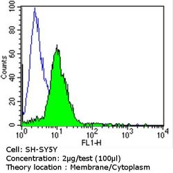
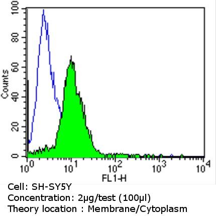
- Flow cytometry analysis of Neurotrophin Receptor /NGF-Receptor (p75NGFR) in SH-SY5Y cells (green) compared to an isotype control (blue). Cells were harvested, adjusted to a concentration of 1-5x10^6 cells/mL, fixed with 2% paraformaldehyde and washed with PBS. Cells were blocked with a 2% solution of BSA-PBS for 30 min at room temperature and incubated with a Neurotrophin Receptor /NGF-Receptor (p75NGFR) monoclonal antibody (Product # MA5-13314) at a dilution of 2 µg/test for 60 min at room temperature. Cells were then incubated for 40 min at room temperature in the dark using a Dylight 488-conjugated goat anti-mouse IgG (H+L) secondary antibody and re-suspended in PBS for FACS analysis.
- Submitted by
- Invitrogen Antibodies (provider)
- Main image

- Experimental details
- Flow cytometry analysis of Neurotrophin Receptor /NGF-Receptor (p75NGFR) in SW480 cells (green) compared to an isotype control (blue). Cells were harvested, adjusted to a concentration of 1-5x10^6 cells/mL, fixed with 2% paraformaldehyde and washed with PBS. Cells were blocked with a 2% solution of BSA-PBS for 30 min at room temperature and incubated with a Neurotrophin Receptor /NGF-Receptor (p75NGFR) monoclonal antibody (Product # MA5-13314) at a dilution of 2 µg/test for 60 min at room temperature. Cells were then incubated for 40 min at room temperature in the dark using a Dylight 488-conjugated goat anti-mouse IgG (H+L) secondary antibody and re-suspended in PBS for FACS analysis.

Supportive validation
- Submitted by
- Invitrogen Antibodies (provider)
- Main image
- Experimental details
- NULL
- Submitted by
- Invitrogen Antibodies (provider)
- Main image
- Experimental details
- NULL
- Submitted by
- Invitrogen Antibodies (provider)
- Main image
- Experimental details
- NULL

- Submitted by
- Invitrogen Antibodies (provider)
- Main image
- Experimental details
- Fig 1 MACS reduces cell stress and improves live cell yield while maintaining an equivalent CD271-/CD133+ sorting efficiency as FACS. (A) Representative immunofluorescence images from one NPC line demonstrating a reduction in CD271+ cells and an enrichment of CD133+ positive cells following both FACS and MACS compared to unsorted cells. Cell nuclei are labelled with DAPI. Scale bar = 100mum, N = 6. (B) Combined flow cytometry analysis of percent CD271+, CD271- and CD271-/CD133+ cells from 100% live cells (DAPI negative population) comparing unsorted, FACS and MACS conditions. (C) Flow cytometry analysis in B presented as individual cell lines. (D) Reduction in CD271+ cell variability following MACS compared to unsorted cell s . **p
