 Explore
 Validate
 Learn
Learn Western blot
Antibody data
- Antibody Data
- Antigen structure
- References [0]
- Comments [0]
- Validations
- Western blot [4]
- Immunocytochemistry [3]
- Immunohistochemistry [2]
Submit
Validation data
Reference
Comment
Report error
- Product number
- PA5-27656 - Provider product page

- Provider
- Invitrogen Antibodies
- Product name
- NGFR Polyclonal Antibody
- Antibody type
- Polyclonal
- Antigen
- Recombinant protein fragment
- Description
- Recommended positive controls: NT2D1, PC-3, U87-MG, PC-12, mouse brain, rat brain. Predicted reactivity: Mouse (92%), Rat (92%), Bovine (97%). Store product as a concentrated solution. Centrifuge briefly prior to opening the vial.
- Reactivity
- Human, Mouse, Rat
- Host
- Rabbit
- Isotype
- IgG
- Vial size
- 100 µL
- Concentration
- 0.43 mg/mL
- Storage
- Store at 4°C short term. For long term storage, store at -20°C, avoiding freeze/thaw cycles.
No comments: Submit comment
Supportive validation
- Submitted by
- Invitrogen Antibodies (provider)
- Main image

- Experimental details
- Western blot analysis of NGFR/p75 NGF Receptor using 30 µg of A) NT2D1 (B) PC-3 and C) U87-MG lysate. Samples were loaded onto a 7.5% SDS-PAGE gel and probed with a NGFR/p75 NGF Receptor polyclonal antibody (Product # PA5-27656) at a dilution of 1:2000.

- Submitted by
- Invitrogen Antibodies (provider)
- Main image
- Experimental details
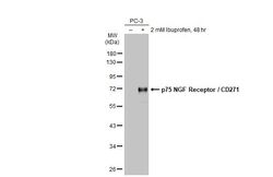
- Western Blot using NGFR Polyclonal Antibody (Product # PA5-27656). Untreated (–) and treated (+) PC-3 whole cell extracts (30 µg) were separated by 10% SDS-PAGE, and the membrane was blotted with p75 NGF Receptor/CD271 antibody NGFR Polyclonal Antibody (Product # PA5-27656) diluted at 1:1,000. The HRP-conjugated anti-rabbit IgG antibody was used to detect the primary antibody.

- Submitted by
- Invitrogen Antibodies (provider)
- Main image
- Experimental details
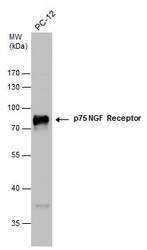
- Western Blot analysis of NGFR was performed by separating 30 µg of whole cell extracts by 10% SDS-PAGE. Proteins were transferred to a membrane and probed with a NGFR Polyclonal Antibody (Product # PA5-27656) at a dilution of 1:2000.

- Submitted by
- Invitrogen Antibodies (provider)
- Main image
- Experimental details
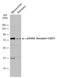
- Western blot analysis of NGFR was performed by separating 50 µg of various tissue extracts by 10% SDS-PAGE. Proteins were transferred to a membrane and probed with a NGFR Polyclonal Antibody (Product # PA5-27656) at a dilution of 1:2000. The HRP-conjugated anti-rabbit IgG antibody was used to detect the primary antibody.

Supportive validation
- Submitted by
- Invitrogen Antibodies (provider)
- Main image

- Experimental details
- Immunocytochemistry-Immunofluorescence analysis of NGFR was performed in Culture rat E18 primary cortical neuron, DIV 8. Cells fixed in 4% paraformaldehyde at RT for 15 min. Green: NGFR Polyclonal Antibody (Product # PA5-27656) diluted at 1:250. Red: beta Tubulin 3/ TUJ1, stained by beta Tubulin 3/ TUJ1 antibody. Blue: Fluoroshield with DAPI.

- Submitted by
- Invitrogen Antibodies (provider)
- Main image

- Experimental details
- NGFR Polyclonal Antibody detects p75 NGF Receptor / CD271 protein by immunofluorescent analysis. Sample: DIV10 rat E18 primary cortical neuron cells were fixed in 4% paraformaldehyde at RT for 15 min. Green: p75 NGF Receptor / CD271 stained by NGFR Polyclonal Antibody (Product # PA5-27656) diluted at 1:500. Red: Tau, stained by Phospho-Tau (Ser262) Polyclonal Antibody [GT287]diluted at 1:500. Blue: Fluoroshield with DAPI .

- Submitted by
- Invitrogen Antibodies (provider)
- Main image

- Experimental details
- Immunocytochemistry-Immunofluorescence analysis of NGFR was performed in Culture rat E18 primary cortical neuron, DIV 8. Cells fixed in 4% paraformaldehyde at RT for 15 min. Green: NGFR Polyclonal Antibody (Product # PA5-27656) diluted at 1:250. Red: beta Tubulin 3/ TUJ1, stained by beta Tubulin 3/ TUJ1 antibody. Blue: Fluoroshield with DAPI.

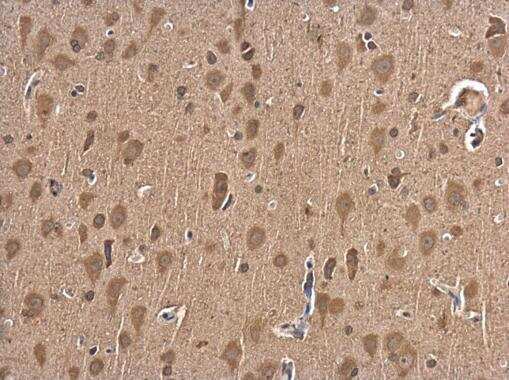
Supportive validation
- Submitted by
- Invitrogen Antibodies (provider)
- Main image
- Experimental details
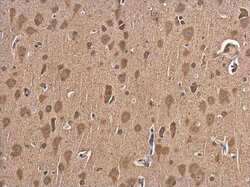
- Immunohistochemistry (Paraffin) analysis of NGFR was performed in paraffin-embedded rat brain tissue using NGFR Polyclonal Antibody (Product # PA5-27656) at a dilution of 1:500.
- Submitted by
- Invitrogen Antibodies (provider)
- Main image

- Experimental details
- Immunohistochemistry (Frozen) analysis of NGFR was performed in frozen sectioned E13.5 Rat brain tissue using NGFR Polyclonal Antibody (Product # PA5-27656) at a dilution of 1:250 (Green). Red: beta Tubulin 3/ TUJ1, a mature neuron marker, stained by beta Tubulin 3/ TUJ1 antibody diluted at 1:500. Blue: Fluoroshield with DAPI.
